Design, Synthesis, and Mechanistic Studies of ( R )-3-Amino-5,5-difluorocyclohex-1-ene-1-carboxylic Acid as an Inactivator of Human Ornithine Aminotransferase.
Devitt, A.N., Vargas, A.L., Zhu, W., Des Soye, B.J., Butun, F.A., Alt, T., Kaley, N., Ferreira, G.M., Moran, G.R., Kelleher, N.L., Liu, D., Silverman, R.B.(2024) ACS Chem Biol 19: 1066-1081
- PubMed: 38630468 Search on PubMed
- DOI: https://doi.org/10.1021/acschembio.4c00022
- Primary Citation Related Structures:
8V9M - PubMed Abstract:
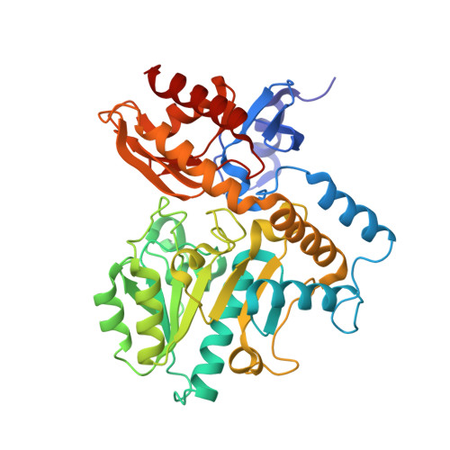
Human ornithine aminotransferase ( h OAT), a pyridoxal 5'-phosphate (PLP)-dependent enzyme, has been shown to play an essential role in the metabolic reprogramming and progression of hepatocellular carcinoma (HCC). HCC accounts for approximately 75% of primary liver cancers and is within the top three causes of cancer death worldwide. As a result of treatment limitations, the overall 5-year survival rate for all patients with HCC is under 20%. The prevalence of HCC necessitates continued development of novel and effective treatment methods. In recent years, the therapeutic potential of selective inactivation of h OAT has been demonstrated for the treatment of HCC. Inspired by previous increased selectivity for h OAT by the expansion of the cyclopentene ring scaffold to a cyclohexene, we designed, synthesized, and evaluated a series of novel fluorinated cyclohexene analogues and identified (R)-3-amino-5,5-difluorocyclohex-1-ene-1-carboxylic acid as a time-dependent inhibitor of h OAT. Structural and mechanistic studies have elucidated the mechanism of inactivation of h OAT by 5 , resulting in a PLP-inactivator adduct tightly bound to the active site of the enzyme. Intact protein mass spectrometry, 19 F NMR spectroscopy, transient state kinetic studies, and X-ray crystallography were used to determine the structure of the final adduct and elucidate the mechanisms of inactivation. Interestingly, despite the highly electrophilic intermediate species conferred by fluorine and structural evidence of solvent accessibility in the h OAT active site, Lys292 and water did not participate in nucleophilic addition during the inactivation mechanism of h OAT by 5 . Instead, rapid aromatization to yield the final adduct was favored.
- Department of Chemistry, Chemistry of Life Processes Institute, and Center for Developmental Therapeutics, Northwestern University, Evanston, Illinois 60208, United States.
Organizational Affiliation: