8V5L | pdb_00008v5l

Structure of the Varicella Zoster Virus (VZV) gI binding domain of glycoprotein E (gE) in complex with human Fab 1A2 and 1E12


Experimental Data Snapshot

  • Method: X-RAY DIFFRACTION
  • Resolution: 3.09 Å
  • R-Value Free: 
    0.269 (Depositor), 0.262 (DCC) 
  • R-Value Work: 
    0.233 (Depositor), 0.230 (DCC) 
  • R-Value Observed: 
    0.236 (Depositor) 

Starting Model: experimental
View more details

wwPDB Validation 3D Report Full Report

Validation slider image for 8V5L

This is version 1.1 of the entry. See complete history

Literature

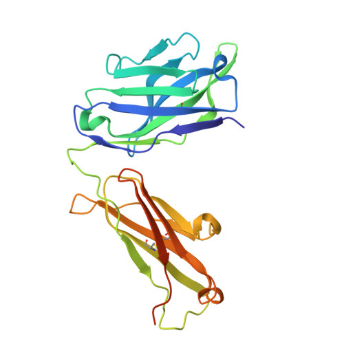
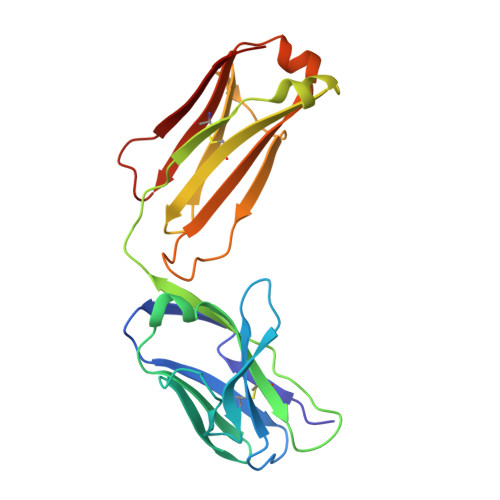
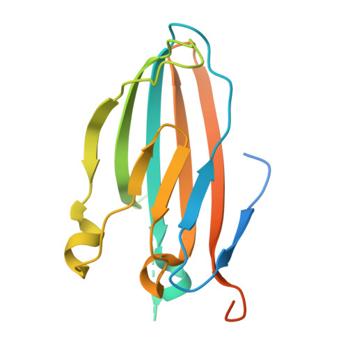
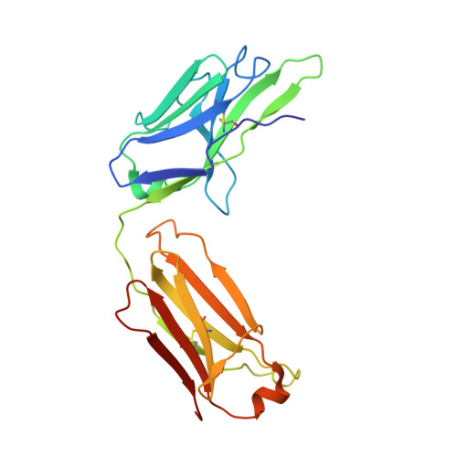
Structures of the Varicella Zoster Virus Glycoprotein E and Epitope Mapping of Vaccine-Elicited Antibodies.

Harshbarger, W.D.Holzapfel, G.Seraj, N.Tian, S.Chesterman, C.Fu, Z.Pan, Y.Harelson, C.Peng, D.Huang, Y.Chandramouli, S.Malito, E.Bottomley, M.J.Williams, J.

(2024) Vaccines (Basel) 12

  • DOI: https://doi.org/10.3390/vaccines12101111
  • Primary Citation Related Structures: 
    8V5L, 8V5P, 8V5Q, 8V5S

  • PubMed Abstract: 

    Background: Varicella zoster virus (VZV) is the causative agent for chickenpox and herpes zoster (HZ, shingles). HZ is a debilitating disease affecting elderly and immunocompromised populations. Glycoprotein E (gE) is indispensable for viral replication and cell-to-cell spread and is the primary target for anti-VZV antibodies. Importantly, gE is the sole antigen in Shingrix, a highly efficacious, AS01 B -adjuvanted vaccine approved in multiple countries for the prevention of HZ, yet the three-dimensional (3D) structure of gE remains elusive. Objectives : We sought to determine the structure of VZV gE and to understand in detail its interactions with neutralizing antibodies. Methods : We used X-ray crystallography and cryo-electron microscopy to elucidate structures of gE bound by recombinant Fabs of antibodies previously elicited through vaccination with Zostavax, a live, attenuated vaccine. Results : The 3D structures resolve distinct central and C-terminal antigenic domains, presenting an array of diverse conformational epitopes. The central domain has two beta-sheets and two alpha helices, including an IgG-like fold. The C-terminal domain exhibits 3 beta-sheets and an Ig-like fold and high structural similarity to HSV1 gE. Conclusions : gE from VZV-infected cells elicits a human antibody response with a preference for the gI binding domain of gE. These results yield insights to VZV gE structure and immunogenicity, provide a framework for future studies, and may guide the design of additional herpesvirus vaccine antigens. Teaser: Structures of varicella zoster virus glycoprotein E reveal distinct antigenic domains and define epitopes for vaccine-elicited human antibodies.


  • Organizational Affiliation
    • GSK, Rockville, MD 20850, USA.

Macromolecule Content 

  • Total Structure Weight: 123.59 kDa 
  • Atom Count: 7,752 
  • Modeled Residue Count: 1,010 
  • Deposited Residue Count: 1,127 
  • Unique protein chains: 5

Macromolecules

Find similar proteins by:|  3D Structure
Entity ID: 1
MoleculeChains  Sequence LengthOrganismDetailsImage
Fab 1A2 Heavy ChainA [auth H]243Homo sapiensMutation(s): 0 
Find similar proteins by:|  3D Structure
Entity ID: 2
MoleculeChains  Sequence LengthOrganismDetailsImage
Fab 1A2 Light ChainB [auth L]214Homo sapiensMutation(s): 0 
Find similar proteins by:|  3D Structure
Entity ID: 3
MoleculeChains  Sequence LengthOrganismDetailsImage
Envelope glycoprotein EC [auth A]203Human alphaherpesvirus 3Mutation(s): 0 
UniProt
Find proteins for A6XEF7 (Human herpesvirus 3)
Explore A6XEF7 
Go to UniProtKB:  A6XEF7
Entity Groups
Sequence Clusters30% Identity50% Identity70% Identity90% Identity95% Identity100% Identity
UniProt GroupA6XEF7
Sequence Annotations
Expand
Reference Sequence
Find similar proteins by:|  3D Structure
Entity ID: 4
MoleculeChains  Sequence LengthOrganismDetailsImage
Fab 1E12 Heavy ChainD [auth B]248Homo sapiensMutation(s): 0 
Find similar proteins by:|  3D Structure
Entity ID: 5
MoleculeChains  Sequence LengthOrganismDetailsImage
Fab 1E12 Light ChainE [auth C]219Homo sapiensMutation(s): 0 

Experimental Data & Validation

Experimental Data

  • Method: X-RAY DIFFRACTION
  • Resolution: 3.09 Å
  • R-Value Free:  0.269 (Depositor), 0.262 (DCC) 
  • R-Value Work:  0.233 (Depositor), 0.230 (DCC) 
  • R-Value Observed: 0.236 (Depositor) 
Space Group: C 1 2 1
Unit Cell:
Length ( Å )Angle ( ˚ )
a = 218.776α = 90
b = 63.493β = 99.96
c = 96.484γ = 90
Software Package:
Software NamePurpose
PHENIXrefinement
PHENIXmodel building
autoPROCdata reduction
PHASERphasing
autoPROCdata scaling

Structure Validation

View Full Validation Report


Entry History 

& Funding Information

Deposition Data


Funding OrganizationLocationGrant Number
Other privateUnited States--

Revision History  (Full details and data files)

  • Version 1.0: 2024-10-09
    Type: Initial release
  • Version 1.1: 2024-11-13
    Changes: Database references