8V5K | pdb_00008v5k

Structure of the Human Respirovirus 3 Fusion Protein Bound to Camelid Nanobodies 4C03 and 4C06


Experimental Data Snapshot

  • Method: ELECTRON MICROSCOPY
  • Resolution: 2.60 Å
  • Aggregation State: PARTICLE 
  • Reconstruction Method: SINGLE PARTICLE 

wwPDB Validation 3D Report Full Report

Validation slider image for 8V5K

This is version 1.2 of the entry. See complete history

Literature

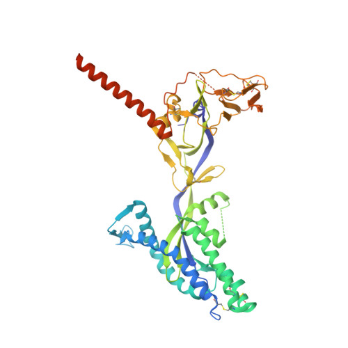
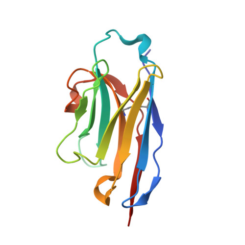
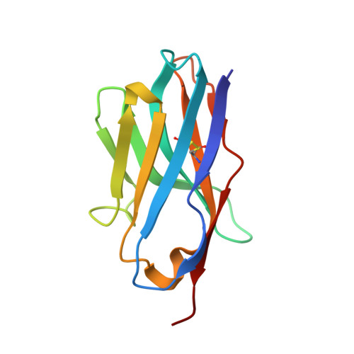
Structural basis for potent neutralization of human respirovirus type 3 by protective single-domain camelid antibodies.

Johnson, N.V.van Scherpenzeel, R.C.Bakkers, M.J.G.Ramamohan, A.R.van Overveld, D.Le, L.Langedijk, J.P.M.Kolkman, J.A.McLellan, J.S.

(2024) Nat Commun 15: 5458-5458

  • DOI: https://doi.org/10.1038/s41467-024-49757-1
  • Primary Citation Related Structures: 
    8V5K, 8V62

  • PubMed Abstract: 

    Respirovirus 3 is a leading cause of severe acute respiratory infections in vulnerable human populations. Entry into host cells is facilitated by the attachment glycoprotein and the fusion glycoprotein (F). Because of its crucial role, F represents an attractive therapeutic target. Here, we identify 13 F-directed heavy-chain-only antibody fragments that neutralize recombinant respirovirus 3. High-resolution cryo-EM structures of antibody fragments bound to the prefusion conformation of F reveal three distinct, previously uncharacterized epitopes. All three antibody fragments bind quaternary epitopes on F, suggesting mechanisms for neutralization that may include stabilization of the prefusion conformation. Studies in cotton rats demonstrate the prophylactic efficacy of these antibody fragments in reducing viral load in the lungs and nasal passages. These data highlight the potential of heavy-chain-only antibody fragments as effective interventions against respirovirus 3 infection and identify neutralizing epitopes that can be targeted for therapeutic development.


  • Organizational Affiliation
    • Department of Molecular Biosciences, The University of Texas at Austin, Austin, TX, 78712, USA.

Macromolecule Content 

  • Total Structure Weight: 245.4 kDa 
  • Atom Count: 15,336 
  • Modeled Residue Count: 1,980 
  • Deposited Residue Count: 2,217 
  • Unique protein chains: 3

Macromolecules

Find similar proteins by:|  3D Structure
Entity ID: 1
MoleculeChains  Sequence LengthOrganismDetailsImage
Fusion glycoprotein F0A,
D [auth B],
E [auth C]
498Human respirovirus 3Mutation(s): 0 
UniProt
Find proteins for A0A023PFZ0 (Human respirovirus 3)
Explore A0A023PFZ0 
Go to UniProtKB:  A0A023PFZ0
Entity Groups
Sequence Clusters30% Identity50% Identity70% Identity90% Identity95% Identity100% Identity
UniProt GroupA0A023PFZ0
Glycosylation
Glycosylation Sites: 1
Sequence Annotations
Expand
Reference Sequence
Find similar proteins by:|  3D Structure
Entity ID: 2
MoleculeChains  Sequence LengthOrganismDetailsImage
Camelid Nanobody 4C03B [auth D],
F [auth E],
G [auth F]
124Lama glamaMutation(s): 0 
Find similar proteins by:|  3D Structure
Entity ID: 3
MoleculeChains  Sequence LengthOrganismDetailsImage
Camelid Nanobody 4C06C [auth G],
H,
I
117Lama glamaMutation(s): 0 

Experimental Data & Validation

Experimental Data

  • Method: ELECTRON MICROSCOPY
  • Resolution: 2.60 Å
  • Aggregation State: PARTICLE 
  • Reconstruction Method: SINGLE PARTICLE 

Structure Validation

View Full Validation Report


Entry History 

& Funding Information

Deposition Data


Funding OrganizationLocationGrant Number
Not funded--

Revision History  (Full details and data files)

  • Version 1.0: 2024-05-22
    Type: Initial release
  • Version 1.1: 2024-07-10
    Changes: Data collection, Database references
  • Version 1.2: 2024-10-30
    Changes: Data collection, Structure summary