8V5B | pdb_00008v5b

Structure of the oxygen-insensitive NAD(P)H-dependent nitroreductase NfsB_Ec F70A/F108Y in complex with FMN


Experimental Data Snapshot

  • Method: X-RAY DIFFRACTION
  • Resolution: 1.98 Å
  • R-Value Free: 
    0.219 (Depositor), 0.225 (DCC) 
  • R-Value Work: 
    0.171 (Depositor), 0.182 (DCC) 
  • R-Value Observed: 
    0.173 (Depositor) 

Starting Model: experimental
View more details

wwPDB Validation 3D Report Full Report

Validation slider image for 8V5B

Ligand Structure Quality Assessment 


This is version 1.0 of the entry. See complete history

Literature

Structural Evaluation of a Nitroreductase Engineered for Improved Activation of the 5-Nitroimidazole PET Probe SN33623.

Sharrock, A.V.Mumm, J.S.Williams, E.M.Cenas, N.Smaill, J.B.Patterson, A.V.Ackerley, D.F.Bagdziunas, G.Arcus, V.L.

(2024) Int J Mol Sci 25

  • DOI: https://doi.org/10.3390/ijms25126593
  • Primary Citation Related Structures: 
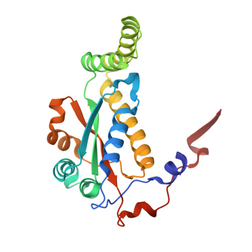
    8V5B

  • PubMed Abstract: 

    Bacterial nitroreductase enzymes capable of activating imaging probes and prodrugs are valuable tools for gene-directed enzyme prodrug therapies and targeted cell ablation models. We recently engineered a nitroreductase ( E. coli NfsB F70A/F108Y) for the substantially enhanced reduction of the 5-nitroimidazole PET-capable probe, SN33623, which permits the theranostic imaging of vectors labeled with oxygen-insensitive bacterial nitroreductases. This mutant enzyme also shows improved activation of the DNA-alkylation prodrugs CB1954 and metronidazole. To elucidate the mechanism behind these enhancements, we resolved the crystal structure of the mutant enzyme to 1.98 Å and compared it to the wild-type enzyme. Structural analysis revealed an expanded substrate access channel and new hydrogen bonding interactions. Additionally, computational modeling of SN33623, CB1954, and metronidazole binding in the active sites of both the mutant and wild-type enzymes revealed key differences in substrate orientations and interactions, with improvements in activity being mirrored by reduced distances between the N5-H of isoalloxazine and the substrate nitro group oxygen in the mutant models. These findings deepen our understanding of nitroreductase substrate specificity and catalytic mechanisms and have potential implications for developing more effective theranostic imaging strategies in cancer treatment.


  • Organizational Affiliation
    • School of Biological Sciences, Victoria University of Wellington, Wellington 6012, New Zealand.

Macromolecule Content 

  • Total Structure Weight: 98.5 kDa 
  • Atom Count: 7,332 
  • Modeled Residue Count: 868 
  • Deposited Residue Count: 868 
  • Unique protein chains: 1

Macromolecules

Find similar proteins by:|  3D Structure
Entity ID: 1
MoleculeChains  Sequence LengthOrganismDetailsImage
Dihydropteridine reductase
A, B, C, D
217Escherichia coliMutation(s): 2 
Gene Names: nfsB
EC: 1 (UniProt), 1.5.1.34 (UniProt)
UniProt
Find proteins for P38489 (Escherichia coli (strain K12))
Explore P38489 
Go to UniProtKB:  P38489
Entity Groups
Sequence Clusters30% Identity50% Identity70% Identity90% Identity95% Identity100% Identity
UniProt GroupP38489
Sequence Annotations
Expand
Reference Sequence

Small Molecules

Ligands 4 Unique
IDChains Name / Formula / InChI Key2D Diagram3D Interactions
P4K

Query on P4K


Download:Ideal Coordinates CCD File
N [auth C]polyethylene glycol
C30 H62 O15
WWPGFZAAWXFBTF-UHFFFAOYSA-N
FMN
(Subject of Investigation/LOI)

Query on FMN


Download:Ideal Coordinates CCD File
E [auth A],
G [auth B],
I [auth C],
O [auth D]
FLAVIN MONONUCLEOTIDE
C17 H21 N4 O9 P
FVTCRASFADXXNN-SCRDCRAPSA-N
ACT

Query on ACT


Download:Ideal Coordinates CCD File
F [auth A]
H [auth B]
J [auth C]
K [auth C]
L [auth C]
F [auth A],
H [auth B],
J [auth C],
K [auth C],
L [auth C],
M [auth C],
P [auth D],
Q [auth D]
ACETATE ION
C2 H3 O2
QTBSBXVTEAMEQO-UHFFFAOYSA-M
CL

Query on CL


Download:Ideal Coordinates CCD File
R [auth D]CHLORIDE ION
Cl
VEXZGXHMUGYJMC-UHFFFAOYSA-M

Experimental Data & Validation

Experimental Data

  • Method: X-RAY DIFFRACTION
  • Resolution: 1.98 Å
  • R-Value Free:  0.219 (Depositor), 0.225 (DCC) 
  • R-Value Work:  0.171 (Depositor), 0.182 (DCC) 
  • R-Value Observed: 0.173 (Depositor) 
Space Group: P 21 21 21
Unit Cell:
Length ( Å )Angle ( ˚ )
a = 87.22α = 90
b = 95.59β = 90
c = 112.31γ = 90
Software Package:
Software NamePurpose
REFMACrefinement
MOSFLMdata reduction
Aimlessdata scaling
PHASERphasing

Structure Validation

View Full Validation Report


Ligand Structure Quality Assessment 


Entry History 

& Funding Information

Deposition Data


Funding OrganizationLocationGrant Number
Marsden FundNew ZealandVUW1902
Other privateNew Zealand--

Revision History  (Full details and data files)

  • Version 1.0: 2024-10-09
    Type: Initial release