8V57

Complex of murine cathepsin K with bound cystatin C inhibitor

  • Classification: HYDROLASE
  • Organism(s): Mus musculus
  • Expression System: Homo sapiens, Escherichia coli
  • Mutation(s): No 

  • Deposited: 2023-11-30 Released: 2024-10-09 
  • Deposition Author(s): Pedersen, L.C., Xu, D.
  • Funding Organization(s): National Institutes of Health/National Institute of Environmental Health Sciences (NIH/NIEHS), National Institutes of Health/National Institute of Dental and Craniofacial Research (NIH/NIDCR), National Institutes of Health/National Institute of Arthritis and Musculoskeletal and Skin Diseases (NIH/NIAMS)

Experimental Data Snapshot

  • Method: X-RAY DIFFRACTION
  • Resolution: 2.75 Å
  • R-Value Free: 0.258 
  • R-Value Work: 0.229 
  • R-Value Observed: 0.230 

Starting Models: experimental
View more details

wwPDB Validation   3D Report Full Report


This is version 1.0 of the entry. See complete history


Literature

Heparan sulfate selectively inhibits the collagenase activity of cathepsin K.

Zhang, X.Luo, Y.Hao, H.Krahn, J.M.Su, G.Dutcher, R.Xu, Y.Liu, J.Pedersen, L.C.Xu, D.

(2024) Matrix Biol 129: 15-28

  • DOI: https://doi.org/10.1016/j.matbio.2024.03.005
  • Primary Citation of Related Structures:  
    8V57, 8V58

  • PubMed Abstract: 

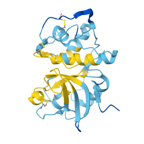
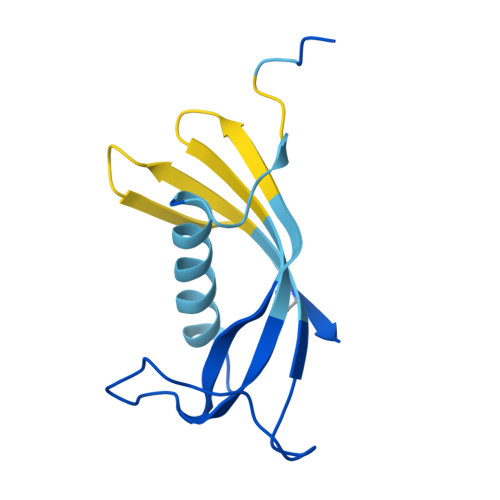
    Cathepsin K (CtsK) is a cysteine protease with potent collagenase activity. CtsK is highly expressed by bone-resorbing osteoclasts and plays an essential role in resorption of bone matrix. Although CtsK is known to bind heparan sulfate (HS), the structural details of the interaction, and how HS regulates the biological functions of CtsK, remains largely unknown. In this report, we discovered that HS is a multifaceted regulator of the structure and function of CtsK. Structurally, HS forms a highly stable complex with CtsK and induces its dimerization. Co-crystal structures of CtsK with bound HS oligosaccharides reveal the location of the HS binding site and suggest how HS may support dimerization. Functionally, HS plays a dual role in regulating the enzymatic activity of CtsK. While it preserves the peptidase activity of CtsK by stabilizing its active conformation, it inhibits the collagenase activity of CtsK in a sulfation level-dependent manner. These opposing effects can be explained by our finding that the HS binding site is remote from the active site, which allows HS to specifically inhibit the collagenase activity without affecting the peptidase activity. At last, we show that structurally defined HS oligosaccharides effectively block osteoclast resorption of bone in vitro without inhibiting osteoclast differentiation, which suggests that HS-based oligosaccharide might be explored as a new class of selective CtsK inhibitor for many diseases involving exaggerated bone resorption.


  • Organizational Affiliation

    Department of Oral Biology, School of Dental Medicine, University at Buffalo, the State University of New York, Buffalo, NY 14214, USA.


Macromolecules
Find similar proteins by:  (by identity cutoff)  |  3D Structure
Entity ID: 1
MoleculeChains Sequence LengthOrganismDetailsImage
Cathepsin K
A, B
217Mus musculusMutation(s): 0 
Gene Names: Ctsk
UniProt & NIH Common Fund Data Resources
Find proteins for P55097 (Mus musculus)
Explore P55097 
Go to UniProtKB:  P55097
IMPC:  MGI:107823
Entity Groups  
Sequence Clusters30% Identity50% Identity70% Identity90% Identity95% Identity100% Identity
UniProt GroupP55097
Glycosylation
Glycosylation Sites: 1Go to GlyGen: P55097-1
Sequence Annotations
Expand
  • Reference Sequence
Find similar proteins by:  (by identity cutoff)  |  3D Structure
Entity ID: 2
MoleculeChains Sequence LengthOrganismDetailsImage
Cystatin-C
C, D
120Mus musculusMutation(s): 0 
Gene Names: Cst3
UniProt & NIH Common Fund Data Resources
Find proteins for P21460 (Mus musculus)
Explore P21460 
Go to UniProtKB:  P21460
IMPC:  MGI:102519
Entity Groups  
Sequence Clusters30% Identity50% Identity70% Identity90% Identity95% Identity100% Identity
UniProt GroupP21460
Sequence Annotations
Expand
  • Reference Sequence
Oligosaccharides

Help

Entity ID: 3
MoleculeChains Length2D Diagram Glycosylation3D Interactions
2-acetamido-2-deoxy-beta-D-glucopyranose-(1-4)-2-acetamido-2-deoxy-beta-D-glucopyranose
E, F
2N-Glycosylation
Small Molecules
Ligands 2 Unique
IDChains Name / Formula / InChI Key2D Diagram3D Interactions
SO4
Query on SO4

Download Ideal Coordinates CCD File 
H [auth A]
I [auth A]
J [auth A]
K [auth A]
L [auth B]
H [auth A],
I [auth A],
J [auth A],
K [auth A],
L [auth B],
M [auth B],
N [auth B],
O [auth B],
P [auth B],
Q [auth C],
R [auth C]
SULFATE ION
O4 S
QAOWNCQODCNURD-UHFFFAOYSA-L
EDO
Query on EDO

Download Ideal Coordinates CCD File 
G [auth A]1,2-ETHANEDIOL
C2 H6 O2
LYCAIKOWRPUZTN-UHFFFAOYSA-N
Experimental Data & Validation

Experimental Data

  • Method: X-RAY DIFFRACTION
  • Resolution: 2.75 Å
  • R-Value Free: 0.258 
  • R-Value Work: 0.229 
  • R-Value Observed: 0.230 
  • Space Group: P 2 2 21
Unit Cell:
Length ( Å )Angle ( ˚ )
a = 92.93α = 90
b = 179.61β = 90
c = 73.352γ = 90
Software Package:
Software NamePurpose
PHENIXrefinement
HKL-2000data reduction
HKL-2000data scaling
PHASERphasing

Structure Validation

View Full Validation Report


Entry History & Funding Information

Deposition Data


Funding OrganizationLocationGrant Number
National Institutes of Health/National Institute of Environmental Health Sciences (NIH/NIEHS)United StatesZIC-ES102645
National Institutes of Health/National Institute of Dental and Craniofacial Research (NIH/NIDCR)United StatesR01DE031273
National Institutes of Health/National Institute of Arthritis and Musculoskeletal and Skin Diseases (NIH/NIAMS)United StatesR01AR070179

Revision History  (Full details and data files)

  • Version 1.0: 2024-10-09
    Type: Initial release