Point mutation in a virus-like capsid drives symmetry reduction to form tetrahedral cages.
Szyszka, T.N., Andreas, M.P., Lie, F., Miller, L.M., Adamson, L.S.R., Fatehi, F., Twarock, R., Draper, B.E., Jarrold, M.F., Giessen, T.W., Lau, Y.H.(2024) Proc Natl Acad Sci U S A 121: e2321260121-e2321260121
- PubMed: 38722807 Search on PubMedSearch on PubMed Central
- DOI: https://doi.org/10.1073/pnas.2321260121
- Primary Citation Related Structures:
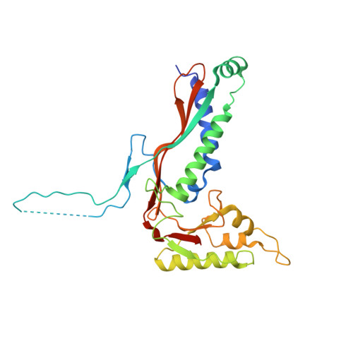
8V4N, 8V4Q - PubMed Abstract:
Protein capsids are a widespread form of compartmentalization in nature. Icosahedral symmetry is ubiquitous in capsids derived from spherical viruses, as this geometry maximizes the internal volume that can be enclosed within. Despite the strong preference for icosahedral symmetry, we show that simple point mutations in a virus-like capsid can drive the assembly of unique symmetry-reduced structures. Starting with the encapsulin from Myxococcus xanthus , a 180-mer bacterial capsid that adopts the well-studied viral HK97 fold, we use mass photometry and native charge detection mass spectrometry to identify a triple histidine point mutant that forms smaller dimorphic assemblies. Using cryoelectron microscopy, we determine the structures of a precedented 60-mer icosahedral assembly and an unexpected 36-mer tetrahedron that features significant geometric rearrangements around a new interaction surface between capsid protomers. We subsequently find that the tetrahedral assembly can be generated by triple-point mutation to various amino acids and that even a single histidine point mutation is sufficient to form tetrahedra. These findings represent a unique example of tetrahedral geometry when surveying all characterized encapsulins, HK97-like capsids, or indeed any virus-derived capsids reported in the Protein Data Bank, revealing the surprising plasticity of capsid self-assembly that can be accessed through minimal changes in the protein sequence.
- School of Chemistry, The University of Sydney, Camperdown, NSW 2006, Australia.
Organizational Affiliation: