8V2A | pdb_00008v2a

Cryo-EM structure of human type I OSM receptor complex: model for assembly core region


Experimental Data Snapshot

  • Method: ELECTRON MICROSCOPY
  • Resolution: 3.59 Å
  • Aggregation State: PARTICLE 
  • Reconstruction Method: SINGLE PARTICLE 

wwPDB Validation 3D Report Full Report

Validation slider image for 8V2A

This is version 1.1 of the entry. See complete history

Literature

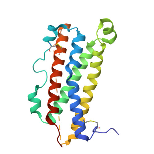
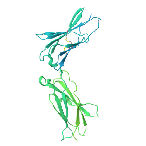
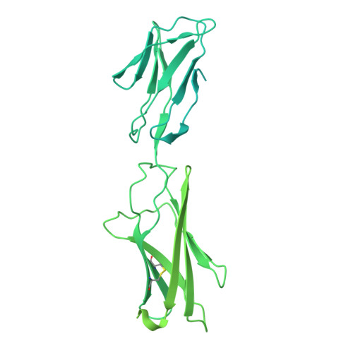
Structures of complete extracellular assemblies of type I and type II Oncostatin M receptor complexes.

Zhou, Y.Stevis, P.E.Cao, J.Ehrlich, G.Jones, J.Rafique, A.Sleeman, M.W.Olson, W.C.Franklin, M.C.

(2024) Nat Commun 15: 9776-9776

  • DOI: https://doi.org/10.1038/s41467-024-54124-1
  • Primary Citation Related Structures: 
    8V29, 8V2A, 8V2B, 8V2C

  • PubMed Abstract: 

    Oncostatin M (OSM) is a unique Interleukin 6 (IL-6) family cytokine that plays pivotal roles in numerous biological events by signaling via two types of receptor complexes. While type I OSM receptor complex is formed by glycoprotein 130 (gp130) heterodimerization with Leukemia Inhibitory Factor receptor (LIFR), type II OSM receptor complex is composed of gp130 and OSM receptor (OSMR). OSM is an important contributor to multiple inflammatory diseases and cancers while OSM inhibition has been shown to be effective at reducing symptoms, making OSM an attractive therapeutic target. Using cryogenic electron microscopy (cryo-EM), we characterize full extracellular assemblies of human type I OSM receptor complex and mouse type II OSM receptor complex. The juxtamembrane domains of both complexes are situated in close proximity due to acute bends of the receptors. The rigid N-terminal extension of OSM contributes to gp130 binding and OSM signaling. Neither glycosylation nor pro-domain cleavage of OSM affects its activity. Mutagenesis identifies multiple OSM and OSMR residues crucial for complex formation and signaling. Our data reveal the structural basis for the assemblies of both type I and type II OSM receptor complexes and provide insights for modulation of OSM signaling in therapeutics.


  • Organizational Affiliation
    • Regeneron Pharmaceuticals, Inc., Tarrytown, NY, 10591, USA. yi.zhou@regeneron.com.

Macromolecule Content 

  • Total Structure Weight: 188.41 kDa 
  • Atom Count: 4,522 
  • Modeled Residue Count: 551 
  • Deposited Residue Count: 1,638 
  • Unique protein chains: 3

Macromolecules

Find similar proteins by:|  3D Structure
Entity ID: 1
MoleculeChains  Sequence LengthOrganismDetailsImage
Oncostatin-M196Homo sapiensMutation(s): 0 
Gene Names: OSM
UniProt & NIH Common Fund Data Resources
Find proteins for P13725 (Homo sapiens)
Explore P13725 
Go to UniProtKB:  P13725
PHAROS:  P13725
GTEx:  ENSG00000099985 
Entity Groups
Sequence Clusters30% Identity50% Identity70% Identity90% Identity95% Identity100% Identity
UniProt GroupP13725
Glycosylation
Glycosylation Sites: 1Go to GlyGen: P13725-1
Sequence Annotations
Expand
Reference Sequence
Find similar proteins by:|  3D Structure
Entity ID: 2
MoleculeChains  Sequence LengthOrganismDetailsImage
Interleukin-6 receptor subunit beta625Homo sapiensMutation(s): 0 
Gene Names: IL6ST
UniProt & NIH Common Fund Data Resources
Find proteins for P40189 (Homo sapiens)
Explore P40189 
Go to UniProtKB:  P40189
PHAROS:  P40189
GTEx:  ENSG00000134352 
Entity Groups
Sequence Clusters30% Identity50% Identity70% Identity90% Identity95% Identity100% Identity
UniProt GroupP40189
Glycosylation
Glycosylation Sites: 3Go to GlyGen: P40189-1
Sequence Annotations
Expand
Reference Sequence
Find similar proteins by:|  3D Structure
Entity ID: 3
MoleculeChains  Sequence LengthOrganismDetailsImage
Leukemia inhibitory factor receptor817Homo sapiensMutation(s): 0 
Gene Names: LIFR
UniProt & NIH Common Fund Data Resources
Find proteins for P42702 (Homo sapiens)
Explore P42702 
Go to UniProtKB:  P42702
PHAROS:  P42702
GTEx:  ENSG00000113594 
Entity Groups
Sequence Clusters30% Identity50% Identity70% Identity90% Identity95% Identity100% Identity
UniProt GroupP42702
Glycosylation
Glycosylation Sites: 3Go to GlyGen: P42702-1
Sequence Annotations
Expand
Reference Sequence

Oligosaccharides

Help  
Entity ID: 4
MoleculeChains Length2D Diagram GlycosylationD Interactions
2-acetamido-2-deoxy-beta-D-glucopyranose-(1-4)-2-acetamido-2-deoxy-beta-D-glucopyranose
D, E, F
2N-Glycosylation
Glycosylation Resources
GlyTouCan: G42666HT
GlyCosmos: G42666HT
GlyGen: G42666HT

Experimental Data & Validation

Experimental Data

  • Method: ELECTRON MICROSCOPY
  • Resolution: 3.59 Å
  • Aggregation State: PARTICLE 
  • Reconstruction Method: SINGLE PARTICLE 
EM Software:
TaskSoftware PackageVersion
MODEL REFINEMENTPHENIX

Structure Validation

View Full Validation Report


Entry History 

& Funding Information

Deposition Data


Funding OrganizationLocationGrant Number
Other private--

Revision History  (Full details and data files)

  • Version 1.0: 2024-11-20
    Type: Initial release
  • Version 1.1: 2025-05-14
    Changes: Data collection