Structural basis for mouse receptor recognition by bat SARS2-like coronaviruses.
Zhang, W., Shi, K., Hsueh, F.C., Mendoza, A., Ye, G., Huang, L., Perlman, S., Aihara, H., Li, F.(2024) Proc Natl Acad Sci U S A 121: e2322600121-e2322600121
- PubMed: 39083418 Search on PubMedSearch on PubMed Central
- DOI: https://doi.org/10.1073/pnas.2322600121
- Primary Citation Related Structures:
8UZE, 8UZF - PubMed Abstract:
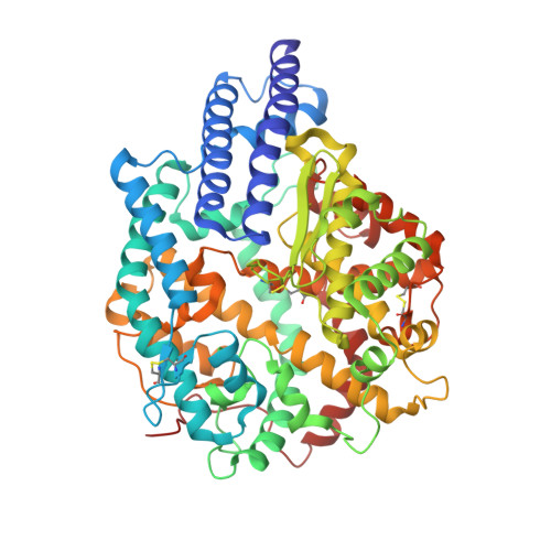
The animal origin of SARS-CoV-2 remains elusive, lacking a plausible evolutionary narrative that may account for its emergence. Its spike protein resembles certain segments of BANAL-236 and RaTG13, two bat coronaviruses considered possible progenitors of SARS-CoV-2. Additionally, its spike contains a furin motif, a common feature of rodent coronaviruses. To explore the possible involvement of rodents in the emergence of SARS-CoV-2 spike, we examined the crystal structures of the spike receptor-binding domains (RBDs) of BANAL-236 and RaTG13 each complexed with mouse receptor ACE2. Both RBDs have residues at positions 493 and 498 that align well with two virus-binding hotspots on mouse ACE2. Our biochemical evidence supports that both BANAL-236 and RaTG13 spikes can use mouse ACE2 as their entry receptor. These findings point to a scenario in which these bat coronaviruses may have coinfected rodents, leading to a recombination of their spike genes and a subsequent acquisition of a furin motif in rodents, culminating in the emergence of SARS-CoV-2.
- Department of Pharmacology, University of Minnesota Medical School, Minneapolis, MN 55455.
Organizational Affiliation: