8UTU

KIF1A[1-393] P305L mutant AMP-PNP bound one and two heads bound states merged, in complex with a microtubule


Experimental Data Snapshot

  • Method: ELECTRON MICROSCOPY
  • Resolution: 3.00 Å
  • Aggregation State: FILAMENT 
  • Reconstruction Method: HELICAL 

wwPDB Validation   3D Report Full Report


This is version 1.1 of the entry. See complete history


Literature

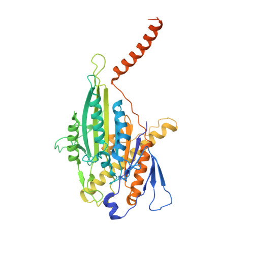
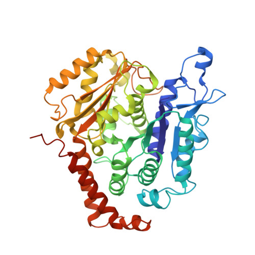
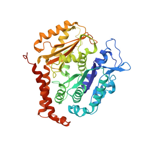
Cryo-EM unveils kinesin KIF1A's processivity mechanism and the impact of its pathogenic variant P305L.

Benoit, M.P.M.H.Rao, L.Asenjo, A.B.Gennerich, A.Sosa, H.

(2024) Nat Commun 15: 5530-5530

  • DOI: https://doi.org/10.1038/s41467-024-48720-4
  • Primary Citation of Related Structures:  
    8UTN, 8UTO, 8UTP, 8UTQ, 8UTR, 8UTS, 8UTT, 8UTU, 8UTV, 8UTW, 8UTY

  • PubMed Abstract: 

    Mutations in the microtubule-associated motor protein KIF1A lead to severe neurological conditions known as KIF1A-associated neurological disorders (KAND). Despite insights into its molecular mechanism, high-resolution structures of KIF1A-microtubule complexes remain undefined. Here, we present 2.7-3.5 Å resolution structures of dimeric microtubule-bound KIF1A, including the pathogenic P305L mutant, across various nucleotide states. Our structures reveal that KIF1A binds microtubules in one- and two-heads-bound configurations, with both heads exhibiting distinct conformations with tight inter-head connection. Notably, KIF1A's class-specific loop 12 (K-loop) forms electrostatic interactions with the C-terminal tails of both α- and β-tubulin. The P305L mutation does not disrupt these interactions but alters loop-12's conformation, impairing strong microtubule-binding. Structure-function analysis reveals the K-loop and head-head coordination as major determinants of KIF1A's superprocessive motility. Our findings advance the understanding of KIF1A's molecular mechanism and provide a basis for developing structure-guided therapeutics against KAND.


  • Organizational Affiliation

    Department of Biochemistry and Gruss-Lipper Biophotonics Center, Albert Einstein College of Medicine, Bronx, NY, 10461, USA. matthieu.benoit@univ-rennes.fr.


Macromolecules
Find similar proteins by:  (by identity cutoff)  |  3D Structure
Entity ID: 1
MoleculeChains Sequence LengthOrganismDetailsImage
Kinesin-like protein KIF1AA [auth K]438Homo sapiensMutation(s): 1 
Gene Names: KIF1A
UniProt & NIH Common Fund Data Resources
Find proteins for Q12756 (Homo sapiens)
Explore Q12756 
Go to UniProtKB:  Q12756
PHAROS:  Q12756
GTEx:  ENSG00000130294 
Entity Groups  
Sequence Clusters30% Identity50% Identity70% Identity90% Identity95% Identity100% Identity
UniProt GroupQ12756
Sequence Annotations
Expand
  • Reference Sequence
Find similar proteins by:  (by identity cutoff)  |  3D Structure
Entity ID: 2
MoleculeChains Sequence LengthOrganismDetailsImage
Tubulin alpha-1B chainB [auth A],
D [auth E]
451Sus scrofaMutation(s): 0 
UniProt
Find proteins for Q2XVP4 (Sus scrofa)
Explore Q2XVP4 
Go to UniProtKB:  Q2XVP4
Entity Groups  
Sequence Clusters30% Identity50% Identity70% Identity90% Identity95% Identity100% Identity
UniProt GroupQ2XVP4
Sequence Annotations
Expand
  • Reference Sequence
Find similar proteins by:  (by identity cutoff)  |  3D Structure
Entity ID: 3
MoleculeChains Sequence LengthOrganismDetailsImage
Tubulin beta-2B chainC [auth B]445Sus scrofaMutation(s): 0 
UniProt
Find proteins for A0A287AGU7 (Sus scrofa)
Explore A0A287AGU7 
Go to UniProtKB:  A0A287AGU7
Entity Groups  
Sequence Clusters30% Identity50% Identity70% Identity90% Identity95% Identity100% Identity
UniProt GroupA0A287AGU7
Sequence Annotations
Expand
  • Reference Sequence
Small Molecules
Ligands 5 Unique
IDChains Name / Formula / InChI Key2D Diagram3D Interactions
TA1
Query on TA1

Download Ideal Coordinates CCD File 
J [auth B]TAXOL
C47 H51 N O14
RCINICONZNJXQF-MZXODVADSA-N
GTP
Query on GTP

Download Ideal Coordinates CCD File 
G [auth A],
K [auth E]
GUANOSINE-5'-TRIPHOSPHATE
C10 H16 N5 O14 P3
XKMLYUALXHKNFT-UUOKFMHZSA-N
ANP
Query on ANP

Download Ideal Coordinates CCD File 
F [auth K]PHOSPHOAMINOPHOSPHONIC ACID-ADENYLATE ESTER
C10 H17 N6 O12 P3
PVKSNHVPLWYQGJ-KQYNXXCUSA-N
GDP
Query on GDP

Download Ideal Coordinates CCD File 
I [auth B]GUANOSINE-5'-DIPHOSPHATE
C10 H15 N5 O11 P2
QGWNDRXFNXRZMB-UUOKFMHZSA-N
MG
Query on MG

Download Ideal Coordinates CCD File 
E [auth K],
H [auth A],
L [auth E]
MAGNESIUM ION
Mg
JLVVSXFLKOJNIY-UHFFFAOYSA-N
Experimental Data & Validation

Experimental Data

  • Method: ELECTRON MICROSCOPY
  • Resolution: 3.00 Å
  • Aggregation State: FILAMENT 
  • Reconstruction Method: HELICAL 

Structure Validation

View Full Validation Report


Entry History & Funding Information

Deposition Data


Funding OrganizationLocationGrant Number
National Institutes of Health/National Institute of General Medical Sciences (NIH/NIGMS)United StatesR01GM113164
National Institutes of Health/National Institute of General Medical Sciences (NIH/NIGMS)United StatesR01GM098469
National Institutes of Health/National Institute of General Medical Sciences (NIH/NIGMS)United StatesR01NS114636
Simons FoundationUnited StatesSF349247
National Institutes of Health/National Institute of General Medical Sciences (NIH/NIGMS)United StatesGM103310
National Institutes of Health/National Institute of General Medical Sciences (NIH/NIGMS)United StatesOD019994

Revision History  (Full details and data files)

  • Version 1.0: 2024-06-12
    Type: Initial release
  • Version 1.1: 2024-07-31
    Changes: Data collection, Database references