8UTA | pdb_00008uta

yjdF riboswitch from R. gauvreauii in complex with proflavine bound to Fab BL3-6 S97N


Experimental Data Snapshot

  • Method: X-RAY DIFFRACTION
  • Resolution: 3.05 Å
  • R-Value Free: 
    0.269 (Depositor), 0.269 (DCC) 
  • R-Value Work: 
    0.224 (Depositor), 0.225 (DCC) 
  • R-Value Observed: 
    0.227 (Depositor) 

Starting Models: experimental
View more details

wwPDB Validation 3D Report Full Report

Validation slider image for 8UTA

Ligand Structure Quality Assessment 


This is version 1.2 of the entry. See complete history

Literature

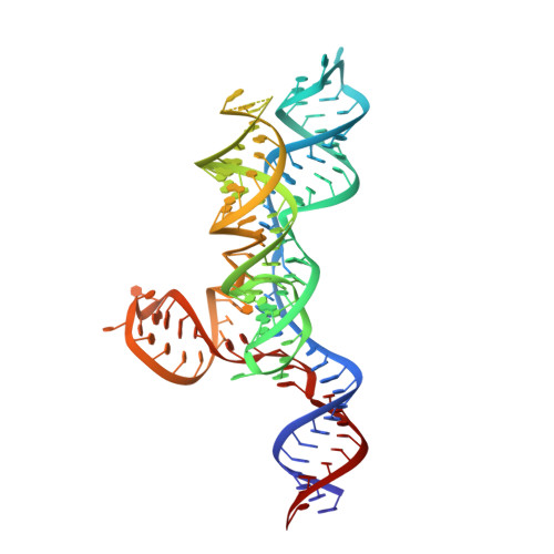
Structural basis for promiscuity in ligand recognition by yjdF riboswitch.

Krochmal, D.Roman, C.Lewicka, A.Shao, Y.Piccirilli, J.A.

(2024) Cell Discov 10: 37-37

Macromolecule Content 

  • Total Structure Weight: 179.17 kDa 
  • Atom Count: 12,024 
  • Modeled Residue Count: 1,131 
  • Deposited Residue Count: 1,152 
  • Unique protein chains: 2
  • Unique nucleic acid chains: 1

Macromolecules


Find similar proteins by:|  3D Structure
Entity ID: 2
MoleculeChains  Sequence LengthOrganismDetailsImage
Fab BL3-6 S97N heavy chainC [auth A],
D [auth H]
233Mus musculusMutation(s): 0 
Find similar proteins by:|  3D Structure
Entity ID: 3
MoleculeChains  Sequence LengthOrganismDetailsImage
Fab BL3-6 S97N light chainE [auth B],
F [auth L]
215Mus musculusMutation(s): 0 
Find similar nucleic acids by:  (by identity cutoff) 
Entity ID: 1
MoleculeChains LengthOrganismImage
yjdF RNA (R. gauvreauii)A [auth N],
B [auth R]
128Ruminococcus gauvreauii
Sequence Annotations
Expand
Reference Sequence

Small Molecules

Ligands 2 Unique
IDChains Name / Formula / InChI Key2D Diagram3D Interactions
PRL
(Subject of Investigation/LOI)

Query on PRL


Download:Ideal Coordinates CCD File
J [auth N],
N [auth R]
PROFLAVIN
C13 H11 N3
WDVSHHCDHLJJJR-UHFFFAOYSA-N
MG

Query on MG


Download:Ideal Coordinates CCD File
G [auth N]
H [auth N]
I [auth N]
K [auth R]
L [auth R]
G [auth N],
H [auth N],
I [auth N],
K [auth R],
L [auth R],
M [auth R]
MAGNESIUM ION
Mg
JLVVSXFLKOJNIY-UHFFFAOYSA-N

Experimental Data & Validation

Experimental Data

  • Method: X-RAY DIFFRACTION
  • Resolution: 3.05 Å
  • R-Value Free:  0.269 (Depositor), 0.269 (DCC) 
  • R-Value Work:  0.224 (Depositor), 0.225 (DCC) 
  • R-Value Observed: 0.227 (Depositor) 
Space Group: P 1
Unit Cell:
Length ( Å )Angle ( ˚ )
a = 63.873α = 87.25
b = 75.138β = 87.99
c = 95.727γ = 87.72
Software Package:
Software NamePurpose
PHENIXrefinement
MOSFLMdata reduction
SCALEPACKdata scaling
PHASERphasing

Structure Validation

View Full Validation Report


Ligand Structure Quality Assessment 


Entry History 

& Funding Information

Deposition Data


Funding OrganizationLocationGrant Number
National Institutes of Health/National Institute of General Medical Sciences (NIH/NIGMS)United StatesR35GM149336

Revision History  (Full details and data files)

  • Version 1.0: 2024-04-10
    Type: Initial release
  • Version 1.1: 2024-05-01
    Changes: Database references
  • Version 1.2: 2024-11-06
    Changes: Structure summary