8UO9

Structure of synaptic vesicle protein 2A in complex with a nanobody


Experimental Data Snapshot

  • Method: ELECTRON MICROSCOPY
  • Resolution: 3.30 Å
  • Aggregation State: PARTICLE 
  • Reconstruction Method: SINGLE PARTICLE 

Starting Model: in silico
View more details

wwPDB Validation   3D Report Full Report


This is version 1.1 of the entry. See complete history


Literature

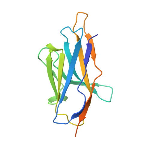
Structures of synaptic vesicle protein 2A and 2B bound to anticonvulsants.

Mittal, A.Martin, M.F.Levin, E.J.Adams, C.Yang, M.Provins, L.Hall, A.Procter, M.Ledecq, M.Hillisch, A.Wolff, C.Gillard, M.Horanyi, P.S.Coleman, J.A.

(2024) Nat Struct Mol Biol 

  • DOI: https://doi.org/10.1038/s41594-024-01335-1
  • Primary Citation of Related Structures:  
    8UO8, 8UO9, 8UOA

  • PubMed Abstract: 

    Epilepsy is a common neurological disorder characterized by abnormal activity of neuronal networks, leading to seizures. The racetam class of anti-seizure medications bind specifically to a membrane protein found in the synaptic vesicles of neurons called synaptic vesicle protein 2 (SV2) A (SV2A). SV2A belongs to an orphan subfamily of the solute carrier 22 organic ion transporter family that also includes SV2B and SV2C. The molecular basis for how anti-seizure medications act on SV2s remains unknown. Here we report cryo-electron microscopy structures of SV2A and SV2B captured in a luminal-occluded conformation complexed with anticonvulsant ligands. The conformation bound by anticonvulsants resembles an inhibited transporter with closed luminal and intracellular gates. Anticonvulsants bind to a highly conserved central site in SV2s. These structures provide blueprints for future drug design and will facilitate future investigations into the biological function of SV2s.


  • Organizational Affiliation

    Department of Structural Biology, University of Pittsburgh, Pittsburgh, PA, USA.


Macromolecules
Find similar proteins by:  (by identity cutoff)  |  3D Structure
Entity ID: 1
MoleculeChains Sequence LengthOrganismDetailsImage
Synaptic vesicle glycoprotein 2A680Homo sapiensMutation(s): 0 
Gene Names: SV2A
Membrane Entity: Yes 
UniProt & NIH Common Fund Data Resources
Find proteins for Q7L0J3 (Homo sapiens)
Explore Q7L0J3 
Go to UniProtKB:  Q7L0J3
PHAROS:  Q7L0J3
GTEx:  ENSG00000159164 
Entity Groups  
Sequence Clusters30% Identity50% Identity70% Identity90% Identity95% Identity100% Identity
UniProt GroupQ7L0J3
Glycosylation
Glycosylation Sites: 3Go to GlyGen: Q7L0J3-1
Sequence Annotations
Expand
  • Reference Sequence
Find similar proteins by:  (by identity cutoff)  |  3D Structure
Entity ID: 2
MoleculeChains Sequence LengthOrganismDetailsImage
Nanobody142Lama glamaMutation(s): 0 
Entity Groups  
Sequence Clusters30% Identity50% Identity70% Identity90% Identity95% Identity100% Identity
Sequence Annotations
Expand
  • Reference Sequence
Oligosaccharides

Help

Entity ID: 3
MoleculeChains Length2D Diagram Glycosylation3D Interactions
beta-D-mannopyranose-(1-4)-2-acetamido-2-deoxy-beta-D-glucopyranose-(1-4)-2-acetamido-2-deoxy-beta-D-glucopyranoseC [auth D]3N-Glycosylation
Glycosylation Resources
GlyTouCan:  G15407YE
GlyCosmos:  G15407YE
GlyGen:  G15407YE
Entity ID: 4
MoleculeChains Length2D Diagram Glycosylation3D Interactions
2-acetamido-2-deoxy-beta-D-glucopyranose-(1-4)-2-acetamido-2-deoxy-beta-D-glucopyranoseD [auth G],
E [auth I]
2N-Glycosylation
Glycosylation Resources
GlyTouCan:  G42666HT
GlyCosmos:  G42666HT
GlyGen:  G42666HT
Small Molecules
Ligands 3 Unique
IDChains Name / Formula / InChI Key2D Diagram3D Interactions
PS1
Query on PS1

Download Ideal Coordinates CCD File 
H [auth A]1,2-DIDECANOYL-SN-GLYCERO-3-[PHOSPHO-L-SERINE]
C26 H49 N O10 P
LRIPXDCMGANCAE-PKTZIBPZSA-M
Y01
Query on Y01

Download Ideal Coordinates CCD File 
G [auth A]CHOLESTEROL HEMISUCCINATE
C31 H50 O4
WLNARFZDISHUGS-MIXBDBMTSA-N
X49 (Subject of Investigation/LOI)
Query on X49

Download Ideal Coordinates CCD File 
F [auth A](4R)-1-{[(4S)-2-(methoxymethyl)-6-(trifluoromethyl)imidazo[2,1-b][1,3,4]thiadiazol-5-yl]methyl}-4-(4,4,4-trifluorobutyl)pyrrolidin-2-one
C16 H18 F6 N4 O2 S
RBBCVRVBYUAJOO-SECBINFHSA-N
Experimental Data & Validation

Experimental Data

  • Method: ELECTRON MICROSCOPY
  • Resolution: 3.30 Å
  • Aggregation State: PARTICLE 
  • Reconstruction Method: SINGLE PARTICLE 
EM Software:
TaskSoftware PackageVersion
MODEL REFINEMENTPHENIX

Structure Validation

View Full Validation Report


Entry History & Funding Information

Deposition Data


Funding OrganizationLocationGrant Number
Brain & Behavior Research FoundationUnited States30153

Revision History  (Full details and data files)

  • Version 1.0: 2024-05-22
    Type: Initial release
  • Version 1.1: 2024-06-26
    Changes: Data collection, Database references, Structure summary