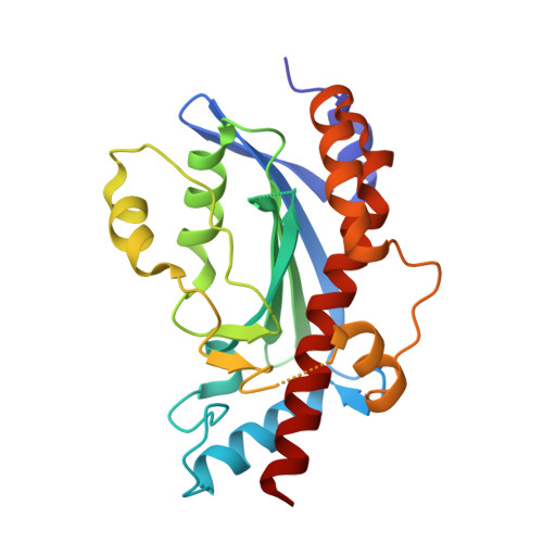
Structural and biochemical characterization of the mitomycin C repair exonuclease MrfB.
Manthei, K.A., Munson, L.M., Nandakumar, J., Simmons, L.A.(2024) Nucleic Acids Res 52: 6347-6359
- PubMed: 38661211 Search on PubMed
- DOI: https://doi.org/10.1093/nar/gkae308
- Primary Citation Related Structures:
8UN9 - PubMed Abstract:
Mitomycin C (MMC) repair factor A (mrfA) and factor B (mrfB), encode a conserved helicase and exonuclease that repair DNA damage in the soil-dwelling bacterium Bacillus subtilis. Here we have focused on the characterization of MrfB, a DEDDh exonuclease in the DnaQ superfamily. We solved the structure of the exonuclease core of MrfB to a resolution of 2.1 Å, in what appears to be an inactive state. In this conformation, a predicted α-helix containing the catalytic DEDDh residue Asp172 adopts a random coil, which moves Asp172 away from the active site and results in the occupancy of only one of the two catalytic Mg2+ ions. We propose that MrfB resides in this inactive state until it interacts with DNA to become activated. By comparing our structure to an AlphaFold prediction as well as other DnaQ-family structures, we located residues hypothesized to be important for exonuclease function. Using exonuclease assays we show that MrfB is a Mg2+-dependent 3'-5' DNA exonuclease. We show that Leu113 aids in coordinating the 3' end of the DNA substrate, and that a basic loop is important for substrate binding. This work provides insight into the function of a recently discovered bacterial exonuclease important for the repair of MMC-induced DNA adducts.
- Department of Molecular, Cellular, and Developmental Biology, University of Michigan, Ann Arbor, MI, USA.
Organizational Affiliation: