8UE2 | pdb_00008ue2

Structure of TREK-1CG*:ML335


Experimental Data Snapshot

  • Method: X-RAY DIFFRACTION
  • Resolution: 3.00 Å
  • R-Value Free: 
    0.292 (Depositor), 0.290 (DCC) 
  • R-Value Work: 
    0.266 (Depositor), 0.271 (DCC) 
  • R-Value Observed: 
    0.268 (Depositor) 

Starting Model: experimental
View more details

wwPDB Validation 3D Report Full Report

Validation slider image for 8UE2

Ligand Structure Quality Assessment 


This is version 1.2 of the entry. See complete history

Literature

Development of covalent chemogenetic K 2P channel activators.

Deal, P.E.Lee, H.Mondal, A.Lolicato, M.Mendonca, P.R.F.Black, H.Jang, S.El-Hilali, X.Bryant, C.Isacoff, E.Y.Renslo, A.R.Minor Jr., D.L.

(2024) Cell Chem Biol 31: 1305-1323.e9

  • DOI: https://doi.org/10.1016/j.chembiol.2024.06.006
  • Primary Citation Related Structures: 
    8UE2, 8UE9, 8UEC, 8UF6

  • PubMed Abstract: 

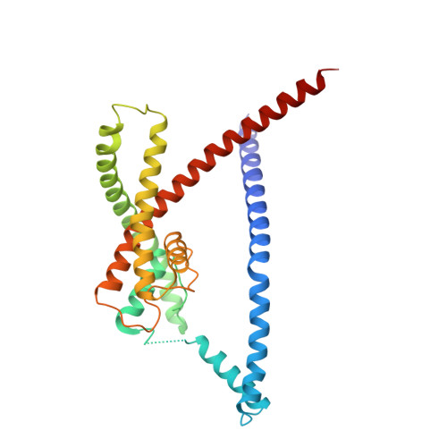
    K 2P potassium channels regulate excitability by affecting cellular resting membrane potential in the brain, cardiovascular system, immune cells, and sensory organs. Despite their important roles in anesthesia, arrhythmia, pain, hypertension, sleep, and migraine, the ability to control K 2P function remains limited. Here, we describe a chemogenetic strategy termed CATKLAMP (covalent activation of TREK family K + channels to clamp membrane potential) that leverages the discovery of a K 2P modulator pocket site that reacts with electrophile-bearing derivatives of a TREK subfamily small-molecule activator, ML335, to activate the channel irreversibly. We show that CATKLAMP can be used to probe fundamental aspects of K 2P function, as a switch to silence neuronal firing, and is applicable to all TREK subfamily members. Together, our findings exemplify a means to alter K 2P channel activity that should facilitate molecular and systems level studies of K 2P function and enable the search for new K 2P modulators.


  • Organizational Affiliation
    • Cardiovascular Research Institute, University of California, San Francisco, San Francisco, CA 93858-2330, USA; Department of Pharmaceutical Chemistry, University of California, San Francisco, San Francisco, CA 93858-2330, USA.

Macromolecule Content 

  • Total Structure Weight: 67.45 kDa 
  • Atom Count: 4,581 
  • Modeled Residue Count: 558 
  • Deposited Residue Count: 574 
  • Unique protein chains: 1

Macromolecules

Find similar proteins by:|  3D Structure
Entity ID: 1
MoleculeChains  Sequence LengthOrganismDetailsImage
Potassium channel subfamily K member 2
A, B
287Mus musculusMutation(s): 14 
Gene Names: Kcnk2
Membrane Entity: Yes 
UniProt & NIH Common Fund Data Resources
Find proteins for P97438 (Mus musculus)
Explore P97438 
Go to UniProtKB:  P97438
IMPC:  MGI:109366
Entity Groups
Sequence Clusters30% Identity50% Identity70% Identity90% Identity95% Identity100% Identity
UniProt GroupP97438
Sequence Annotations
Expand
Reference Sequence

Small Molecules

Ligands 5 Unique
IDChains Name / Formula / InChI Key2D Diagram3D Interactions
Q6F
(Subject of Investigation/LOI)

Query on Q6F


Download:Ideal Coordinates CCD File
AA [auth B],
L [auth A]
N-[(2,4-dichlorophenyl)methyl]-4-[(methylsulfonyl)amino]benzamide
C15 H14 Cl2 N2 O3 S
RDFIQTZRJRVFHK-UHFFFAOYSA-N
R16

Query on R16


Download:Ideal Coordinates CCD File
F [auth A]
G [auth A]
H [auth A]
I [auth A]
J [auth A]
F [auth A],
G [auth A],
H [auth A],
I [auth A],
J [auth A],
M [auth A],
U [auth B],
V [auth B],
W [auth B],
X [auth B],
Y [auth B],
Z [auth B]
HEXADECANE
C16 H34
DCAYPVUWAIABOU-UHFFFAOYSA-N
D10

Query on D10


Download:Ideal Coordinates CCD File
K [auth A]DECANE
C10 H22
DIOQZVSQGTUSAI-UHFFFAOYSA-N
CD

Query on CD


Download:Ideal Coordinates CCD File
N [auth A],
R [auth B],
S [auth B],
T [auth B]
CADMIUM ION
Cd
WLZRMCYVCSSEQC-UHFFFAOYSA-N
K

Query on K


Download:Ideal Coordinates CCD File
C [auth A]
D [auth A]
E [auth A]
O [auth B]
P [auth B]
C [auth A],
D [auth A],
E [auth A],
O [auth B],
P [auth B],
Q [auth B]
POTASSIUM ION
K
NPYPAHLBTDXSSS-UHFFFAOYSA-N

Experimental Data & Validation

Experimental Data

  • Method: X-RAY DIFFRACTION
  • Resolution: 3.00 Å
  • R-Value Free:  0.292 (Depositor), 0.290 (DCC) 
  • R-Value Work:  0.266 (Depositor), 0.271 (DCC) 
  • R-Value Observed: 0.268 (Depositor) 
Space Group: P 21 21 21
Unit Cell:
Length ( Å )Angle ( ˚ )
a = 66.929α = 90
b = 119.655β = 90
c = 128.652γ = 90
Software Package:
Software NamePurpose
PHENIXrefinement
XDSdata reduction
XDSdata scaling
PHASERphasing

Structure Validation

View Full Validation Report


Ligand Structure Quality Assessment 


Entry History 

& Funding Information

Deposition Data


Funding OrganizationLocationGrant Number
National Institutes of Health/National Institute of Mental Health (NIH/NIMH)United StatesR01-MH116278
National Institutes of Health/National Institute of Mental Health (NIH/NIMH)United StatesR01-MH093603

Revision History  (Full details and data files)

  • Version 1.0: 2024-06-26
    Type: Initial release
  • Version 1.1: 2024-11-06
    Changes: Structure summary
  • Version 1.2: 2025-01-08
    Changes: Database references