8UDK

Human Mitochondrial DNA Polymerase gamma R853A Ternary Complex


Experimental Data Snapshot

  • Method: X-RAY DIFFRACTION
  • Resolution: 3.43 Å
  • R-Value Free: 0.234 
  • R-Value Work: 0.198 
  • R-Value Observed: 0.199 

Starting Model: experimental
View more details

wwPDB Validation   3D Report Full Report


Ligand Structure Quality Assessment 


This is version 1.0 of the entry. See complete history


Literature

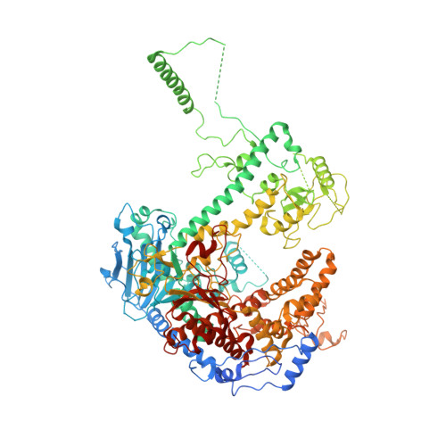
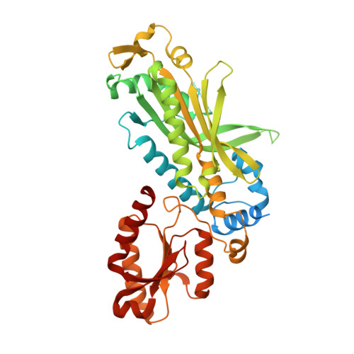
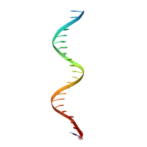
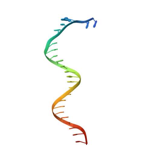
An interaction network in the polymerase active site is a prerequisite for Watson-Crick base pairing in Pol gamma.

Park, J.Herrmann, G.K.Roy, A.Shumate, C.K.Cisneros, G.A.Yin, Y.W.

(2024) Sci Adv 10: eadl3214-eadl3214

  • DOI: https://doi.org/10.1126/sciadv.adl3214
  • Primary Citation of Related Structures:  
    8UDK, 8UDL

  • PubMed Abstract: 

    The replication accuracy of DNA polymerase gamma (Pol γ) is essential for mitochondrial genome integrity. Mutation of human Pol γ arginine-853 has been linked to neurological diseases. Although not a catalytic residue, Pol γ arginine-853 mutants are void of polymerase activity. To identify the structural basis for the disease, we determined a crystal structure of the Pol γ mutant ternary complex with correct incoming nucleotide 2'-deoxycytidine 5'-triphosphate (dCTP). Opposite to the wild type that undergoes open-to-closed conformational changes when bound to a correct nucleotide that is essential for forming a catalytically competent active site, the mutant complex failed to undergo the conformational change, and the dCTP did not base pair with its Watson-Crick complementary templating residue. Our studies revealed that arginine-853 coordinates an interaction network that aligns the 3'-end of primer and dCTP with the catalytic residues. Disruption of the network precludes the formation of Watson-Crick base pairing and closing of the active site, resulting in an inactive polymerase.


  • Organizational Affiliation

    Department of Biochemistry and Molecular Biology, University of Texas Medical Branch, Galveston, TX 77555, USA.


Macromolecules

Find similar proteins by:  (by identity cutoff)  |  3D Structure
Entity ID: 1
MoleculeChains Sequence LengthOrganismDetailsImage
DNA polymerase subunit gamma-11,239Homo sapiensMutation(s): 3 
Gene Names: POLGMDP1POLG1POLGA
EC: 2.7.7.7 (PDB Primary Data), 3.1.11 (PDB Primary Data), 4.2.99 (PDB Primary Data)
UniProt & NIH Common Fund Data Resources
Find proteins for P54098 (Homo sapiens)
Explore P54098 
Go to UniProtKB:  P54098
PHAROS:  P54098
GTEx:  ENSG00000140521 
Entity Groups  
Sequence Clusters30% Identity50% Identity70% Identity90% Identity95% Identity100% Identity
UniProt GroupP54098
Sequence Annotations
Expand
  • Reference Sequence
Find similar proteins by:  (by identity cutoff)  |  3D Structure
Entity ID: 2
MoleculeChains Sequence LengthOrganismDetailsImage
DNA polymerase subunit gamma-2, mitochondrial
B, C
485Homo sapiensMutation(s): 0 
Gene Names: POLG2MTPOLB
EC: 2.7.7.7
UniProt & NIH Common Fund Data Resources
Find proteins for Q9UHN1 (Homo sapiens)
Explore Q9UHN1 
Go to UniProtKB:  Q9UHN1
PHAROS:  Q9UHN1
GTEx:  ENSG00000256525 
Entity Groups  
Sequence Clusters30% Identity50% Identity70% Identity90% Identity95% Identity100% Identity
UniProt GroupQ9UHN1
Sequence Annotations
Expand
  • Reference Sequence

Find similar nucleic acids by:  Sequence   |   3D Structure  

Entity ID: 3
MoleculeChains LengthOrganismImage
DNA (24-MER)D [auth P]24synthetic construct
Sequence Annotations
Expand
  • Reference Sequence
Find similar nucleic acids by:  (by identity cutoff)  |  3D Structure
Entity ID: 4
MoleculeChains LengthOrganismImage
DNA (28-MER)E [auth T]28synthetic construct
Sequence Annotations
Expand
  • Reference Sequence

Find similar nucleic acids by:  Sequence   |   3D Structure  

Entity ID: 5
MoleculeChains LengthOrganismImage
DNA (5'-D(P*AP*AP*GP*GP*GP*CP*CP*TP*AP*TP*AP*AP*AP*A)-3')F [auth D]14synthetic construct
Sequence Annotations
Expand
  • Reference Sequence

Find similar nucleic acids by:  Sequence   |   3D Structure  

Entity ID: 6
MoleculeChains LengthOrganismImage
DNA (5'-D(P*TP*TP*TP*TP*AP*TP*AP*GP*GP*CP*CP*CP*TP*T)-3')G [auth E]14synthetic construct
Sequence Annotations
Expand
  • Reference Sequence
Small Molecules
Ligands 1 Unique
IDChains Name / Formula / InChI Key2D Diagram3D Interactions
DCP (Subject of Investigation/LOI)
Query on DCP

Download Ideal Coordinates CCD File 
H [auth A]2'-DEOXYCYTIDINE-5'-TRIPHOSPHATE
C9 H16 N3 O13 P3
RGWHQCVHVJXOKC-SHYZEUOFSA-N
Experimental Data & Validation

Experimental Data

  • Method: X-RAY DIFFRACTION
  • Resolution: 3.43 Å
  • R-Value Free: 0.234 
  • R-Value Work: 0.198 
  • R-Value Observed: 0.199 
  • Space Group: P 41 21 2
Unit Cell:
Length ( Å )Angle ( ˚ )
a = 215.41α = 90
b = 215.41β = 90
c = 169.966γ = 90
Software Package:
Software NamePurpose
PHENIXrefinement
HKL-3000data reduction
HKL-3000data scaling
PHENIXphasing

Structure Validation

View Full Validation Report


Ligand Structure Quality Assessment 


Entry History & Funding Information

Deposition Data


Funding OrganizationLocationGrant Number
National Institutes of Health/National Institute Of Allergy and Infectious Diseases (NIH/NIAID)United StatesR01AI134611
National Institutes of Health/National Institute of General Medical Sciences (NIH/NIGMS)United StatesR01GM145925

Revision History  (Full details and data files)

  • Version 1.0: 2024-06-05
    Type: Initial release