Moderately thermostable GH1 beta-glucosidases from hyperacidophilic archaeon Cuniculiplasma divulgatum S5.
Khusnutdinova, A.N., Tran, H., Devlekar, S., Distaso, M.A., Kublanov, I.V., Skarina, T., Stogios, P., Savchenko, A., Ferrer, M., Golyshina, O.V., Yakunin, A.F., Golyshin, P.N.(2024) FEMS Microbiol Ecol 100
- PubMed: 39127612 Search on PubMedSearch on PubMed Central
- DOI: https://doi.org/10.1093/femsec/fiae114
- Primary Citation Related Structures:
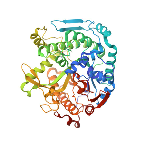
8U7F, 8U7G - PubMed Abstract:
Family GH1 glycosyl hydrolases are ubiquitous in prokaryotes and eukaryotes and are utilized in numerous industrial applications, including bioconversion of lignocelluloses. In this study, hyperacidophilic archaeon Cuniculiplasma divulgatum (S5T=JCM 30642T) was explored as a source of novel carbohydrate-active enzymes. The genome of C. divulgatum encodes three GH1 enzyme candidates, from which CIB12 and CIB13 were heterologously expressed and characterized. Phylogenetic analysis of CIB12 and CIB13 clustered them with β-glucosidases from genuinely thermophilic archaea including Thermoplasma acidophilum, Picrophilus torridus, Sulfolobus solfataricus, Pyrococcus furiosus, and Thermococcus kodakarensis. Purified enzymes showed maximal activities at pH 4.5-6.0 (CIB12) and 4.5-5.5 (CIB13) with optimal temperatures at 50°C, suggesting a high-temperature origin of Cuniculiplasma spp. ancestors. Crystal structures of both enzymes revealed a classical (α/β)8 TIM-barrel fold with the active site located inside the barrel close to the C-termini of β-strands including the catalytic residues Glu204 and Glu388 (CIB12), and Glu204 and Glu385 (CIB13). Both enzymes preferred cellobiose over lactose as substrates and were classified as cellobiohydrolases. Cellobiose addition increased the biomass yield of Cuniculiplasma cultures growing on peptides by 50%, suggesting that the cellobiohydrolases expand the carbon substrate range and hence environmental fitness of Cuniculiplasma.
- Centre for Environmental Biotechnology, School of Environmental and Natural Sciences, Bangor University, Bangor, LL57 2UW, United Kingdom.
Organizational Affiliation: