Deep-branching evolutionary intermediates reveal structural origins of form I rubisco.
Liu, A.K., Kaeser, B., Chen, L., West-Roberts, J., Taylor-Kearney, L.J., Lavy, A., Gunzing, D., Li, W.J., Hammel, M., Nogales, E., Banfield, J.F., Shih, P.M.(2023) Curr Biol 33: 5316-5325.e3
- PubMed: 37979578 Search on PubMed
- DOI: https://doi.org/10.1016/j.cub.2023.10.053
- Primary Citation Related Structures:
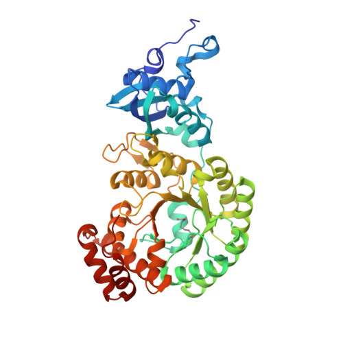
8U66 - PubMed Abstract:
The enzyme rubisco (ribulose-1,5-bisphosphate carboxylase/oxygenase) catalyzes the majority of biological carbon fixation on Earth. Although the vast majority of rubiscos across the tree of life assemble as homo-oligomers, the globally predominant form I enzyme-found in plants, algae, and cyanobacteria-forms a unique hetero-oligomeric complex. The recent discovery of a homo-oligomeric sister group to form I rubisco (named form I') has filled a key gap in our understanding of the enigmatic origins of the form I clade. However, to elucidate the series of molecular events leading to the evolution of form I rubisco, we must examine more distantly related sibling clades to contextualize the molecular features distinguishing form I and form I' rubiscos. Here, we present a comparative structural study retracing the evolutionary history of rubisco that reveals a complex structural trajectory leading to the ultimate hetero-oligomerization of the form I clade. We structurally characterize the oligomeric states of deep-branching form Iα and I'' rubiscos recently discovered from metagenomes, which represent key evolutionary intermediates preceding the form I clade. We further solve the structure of form I'' rubisco, revealing the molecular determinants that likely primed the enzyme core for the transition from a homo-oligomer to a hetero-oligomer. Our findings yield new insight into the evolutionary trajectory underpinning the adoption and entrenchment of the prevalent assembly of form I rubisco, providing additional context when viewing the enzyme family through the broader lens of protein evolution.
- Department of Plant and Microbial Biology, University of California, Berkeley, Berkeley, CA 94720, USA; Environmental Genomics and Systems Biology Division, Lawrence Berkeley National Laboratory, Berkeley, CA 94720, USA; Feedstocks Division, Joint BioEnergy Institute, Emeryville, CA, USA; Biochemistry, Molecular, Cellular and Developmental Biology Graduate Group, University of California, Davis, Davis, CA 95616, USA.
Organizational Affiliation: