8U3L | pdb_00008u3l

TRPV1 in nanodisc bound with empty vanilloid binding pocket at 25C


Experimental Data Snapshot

  • Method: ELECTRON MICROSCOPY
  • Resolution: 3.70 Å
  • Aggregation State: PARTICLE 
  • Reconstruction Method: SINGLE PARTICLE 

wwPDB Validation 3D Report Full Report

Validation slider image for 8U3L

This is version 1.4 of the entry. See complete history

Literature

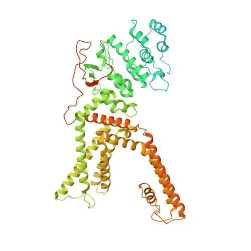
Structural basis of TRPV1 modulation by endogenous bioactive lipids.

Arnold, W.R.Mancino, A.Moss 3rd, F.R.Frost, A.Julius, D.Cheng, Y.

(2024) Nat Struct Mol Biol 31: 1377-1385

  • DOI: https://doi.org/10.1038/s41594-024-01299-2
  • Primary Citation Related Structures: 
    8T0C, 8T0E, 8T0Y, 8T10, 8T3L, 8T3M, 8U2Z, 8U30, 8U3A, 8U3C, 8U3J, 8U3L, 8U43, 8U4D

  • PubMed Abstract: 

    TRP ion channels are modulated by phosphoinositide lipids, but the underlying structural mechanisms remain unclear. The capsaicin- and heat-activated receptor, TRPV1, has served as a model for deciphering lipid modulation, which is relevant to understanding how pro-algesic agents enhance channel activity in the setting of inflammatory pain. Identification of a pocket within the TRPV1 transmembrane core has provided initial clues as to how phosphoinositide lipids bind to and regulate the channel. Here we show that this regulatory pocket in rat TRPV1 can accommodate diverse lipid species, including the inflammatory lipid lysophosphatidic acid, whose actions are determined by their specific modes of binding. Furthermore, we show that an empty-pocket channel lacking an endogenous phosphoinositide lipid assumes an agonist-like state, even at low temperature, substantiating the concept that phosphoinositide lipids serve as negative TRPV1 modulators whose ejection from the binding pocket is a critical step toward activation by thermal or chemical stimuli.


  • Organizational Affiliation
    • Department of Biochemistry and Biophysics, University of California San Francisco, San Francisco, CA, USA.

Macromolecule Content 

  • Total Structure Weight: 373.46 kDa 
  • Atom Count: 17,461 
  • Modeled Residue Count: 2,132 
  • Deposited Residue Count: 3,260 
  • Unique protein chains: 1

Macromolecules

Find similar proteins by:|  3D Structure
Entity ID: 1
MoleculeChains  Sequence LengthOrganismDetailsImage
Transient receptor potential cation channel subfamily V member 1A,
B [auth D],
C [auth B],
D [auth C]
815Rattus norvegicusMutation(s): 0 
Gene Names: Trpv1Vr1Vr1l
Membrane Entity: Yes 
UniProt
Find proteins for O35433 (Rattus norvegicus)
Explore O35433 
Go to UniProtKB:  O35433
Entity Groups
Sequence Clusters30% Identity50% Identity70% Identity90% Identity95% Identity100% Identity
UniProt GroupO35433
Sequence Annotations
Expand
Reference Sequence

Experimental Data & Validation

Experimental Data

  • Method: ELECTRON MICROSCOPY
  • Resolution: 3.70 Å
  • Aggregation State: PARTICLE 
  • Reconstruction Method: SINGLE PARTICLE 
EM Software:
TaskSoftware PackageVersion
MODEL REFINEMENTPHENIX

Structure Validation

View Full Validation Report


Entry History 

& Funding Information

Deposition Data


Funding OrganizationLocationGrant Number
National Institutes of Health/National Institute of General Medical Sciences (NIH/NIGMS)United States--
Howard Hughes Medical Institute (HHMI)United States--

Revision History  (Full details and data files)

  • Version 1.0: 2024-05-08
    Type: Initial release
  • Version 1.1: 2024-05-15
    Changes: Database references
  • Version 1.2: 2024-09-25
    Changes: Data collection, Database references
  • Version 1.3: 2024-10-02
    Changes: Data collection, Database references
  • Version 1.4: 2025-06-04
    Changes: Data collection, Structure summary