


Bacterial fluorescent protein
Gully, B.S., Rossjohn, J.To be published.
Experimental Data Snapshot
Starting Model: experimental
View more details
Entity ID: 1 | |||||
|---|---|---|---|---|---|
| Molecule | Chains | Sequence Length | Organism | Details | Image |
| Bacterial fluorescent protein alpha | A [auth B], C [auth K] | 164 | Ceramium secundatum | Mutation(s): 0 Gene Names: cpeA |  |
UniProt | |||||
Entity Groups | |||||
| Sequence Clusters | 30% Identity50% Identity70% Identity90% Identity95% Identity100% Identity | ||||
| UniProt Group | Q1AH74 | ||||
Sequence AnnotationsExpand | |||||
Reference Sequence | |||||
Entity ID: 2 | |||||
|---|---|---|---|---|---|
| Molecule | Chains | Sequence Length | Organism | Details | Image |
| Bacterial fluorescent protein beta | B [auth C], D [auth L] | 176 | Ceramium secundatum | Mutation(s): 0 Gene Names: cpeB | 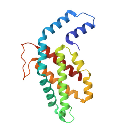 |
UniProt | |||||
Entity Groups | |||||
| Sequence Clusters | 30% Identity50% Identity70% Identity90% Identity95% Identity100% Identity | ||||
| UniProt Group | Q1AH60 | ||||
Sequence AnnotationsExpand | |||||
Reference Sequence | |||||
| Ligands 2 Unique | |||||
|---|---|---|---|---|---|
| ID | Chains | Name / Formula / InChI Key | 2D Diagram | 3D Interactions | |
| PUB Download:Ideal Coordinates CCD File | G [auth C], L | PHYCOUROBILIN C33 H42 N4 O6 KDCCOOGTVSRCHX-YYVBKQGDSA-N |  | ||
| CYC Download:Ideal Coordinates CCD File | E [auth B] F [auth B] H [auth C] I [auth C] J [auth K] | PHYCOCYANOBILIN C33 H40 N4 O6 VXTXPYZGDQPMHK-GMXXPEQVSA-N |  | ||
| Length ( Å ) | Angle ( ˚ ) |
|---|---|
| a = 186.61 | α = 90 |
| b = 186.61 | β = 90 |
| c = 59.42 | γ = 120 |
| Software Name | Purpose |
|---|---|
| PHENIX | refinement |
| MOSFLM | data reduction |
| SCALA | data scaling |
| PHASER | phasing |
| Funding Organization | Location | Grant Number |
|---|---|---|
| Australian Research Council (ARC) | Australia | DP230102073 |


