8TZX

Ternary complex structure of Cereblon-DDB1 bound to WIZ(ZF7) and the molecular glue dWIZ-1


Experimental Data Snapshot

  • Method: X-RAY DIFFRACTION
  • Resolution: 3.15 Å
  • R-Value Free: 0.252 
  • R-Value Work: 0.213 
  • R-Value Observed: 0.215 

Starting Model: experimental
View more details

wwPDB Validation   3D Report Full Report


Ligand Structure Quality Assessment 


This is version 1.1 of the entry. See complete history


Literature

A molecular glue degrader of the WIZ transcription factor for fetal hemoglobin induction.

Ting, P.Y.Borikar, S.Kerrigan, J.R.Thomsen, N.M.Aghania, E.Hinman, A.E.Reyes, A.Pizzato, N.Fodor, B.D.Wu, F.Belew, M.S.Mao, X.Wang, J.Chitnis, S.Niu, W.Hachey, A.Cobb, J.S.Savage, N.A.Burke, A.Paulk, J.Dovala, D.Lin, J.Clifton, M.C.Ornelas, E.Ma, X.Ware, N.F.Sanchez, C.C.Taraszka, J.Terranova, R.Knehr, J.Altorfer, M.Barnes, S.W.Beckwith, R.E.J.Solomon, J.M.Dales, N.A.Patterson, A.W.Wagner, J.Bouwmeester, T.Dranoff, G.Stevenson, S.C.Bradner, J.E.

(2024) Science 385: 91-99

  • DOI: https://doi.org/10.1126/science.adk6129
  • Primary Citation of Related Structures:  
    8TZX

  • PubMed Abstract: 

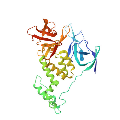
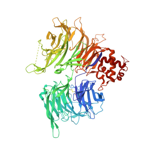
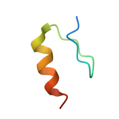
    Sickle cell disease (SCD) is a prevalent, life-threatening condition attributable to a heritable mutation in β-hemoglobin. Therapeutic induction of fetal hemoglobin (HbF) can ameliorate disease complications and has been intently pursued. However, safe and effective small-molecule inducers of HbF remain elusive. We report the discovery of dWIZ-1 and dWIZ-2, molecular glue degraders of the WIZ transcription factor that robustly induce HbF in erythroblasts. Phenotypic screening of a cereblon (CRBN)-biased chemical library revealed WIZ as a previously unknown repressor of HbF. WIZ degradation is mediated by recruitment of WIZ(ZF7) to CRBN by dWIZ-1, as resolved by crystallography of the ternary complex. Pharmacological degradation of WIZ was well tolerated and induced HbF in humanized mice and cynomolgus monkeys. These findings establish WIZ degradation as a globally accessible therapeutic strategy for SCD.


  • Organizational Affiliation

    Novartis Biomedical Research, Cambridge, MA, USA.


Macromolecules
Find similar proteins by:  (by identity cutoff)  |  3D Structure
Entity ID: 1
MoleculeChains Sequence LengthOrganismDetailsImage
Protein cereblon
A, D
373Homo sapiensMutation(s): 0 
Gene Names: CRBN
UniProt & NIH Common Fund Data Resources
Find proteins for Q96SW2 (Homo sapiens)
Explore Q96SW2 
Go to UniProtKB:  Q96SW2
GTEx:  ENSG00000113851 
Entity Groups  
Sequence Clusters30% Identity50% Identity70% Identity90% Identity95% Identity100% Identity
UniProt GroupQ96SW2
Sequence Annotations
Expand
  • Reference Sequence
Find similar proteins by:  (by identity cutoff)  |  3D Structure
Entity ID: 2
MoleculeChains Sequence LengthOrganismDetailsImage
DNA damage-binding protein 1
B, E
836Homo sapiensMutation(s): 0 
Gene Names: DDB1XAP1
UniProt & NIH Common Fund Data Resources
Find proteins for Q16531 (Homo sapiens)
Explore Q16531 
Go to UniProtKB:  Q16531
GTEx:  ENSG00000167986 
Entity Groups  
Sequence Clusters30% Identity50% Identity70% Identity90% Identity95% Identity100% Identity
UniProt GroupQ16531
Sequence Annotations
Expand
  • Reference Sequence
Find similar proteins by:  (by identity cutoff)  |  3D Structure
Entity ID: 3
MoleculeChains Sequence LengthOrganismDetailsImage
Protein Wiz
C, F
30Homo sapiensMutation(s): 0 
Gene Names: WIZZNF803
UniProt & NIH Common Fund Data Resources
Find proteins for O95785 (Homo sapiens)
Explore O95785 
Go to UniProtKB:  O95785
PHAROS:  O95785
GTEx:  ENSG00000011451 
Entity Groups  
Sequence Clusters30% Identity50% Identity70% Identity90% Identity95% Identity100% Identity
UniProt GroupO95785
Sequence Annotations
Expand
  • Reference Sequence
Small Molecules
Ligands 4 Unique
IDChains Name / Formula / InChI Key2D Diagram3D Interactions
U3I (Subject of Investigation/LOI)
Query on U3I

Download Ideal Coordinates CCD File 
H [auth A],
N [auth D]
(3S)-3-(5-{(1R)-1-[(2R)-1-ethylpiperidin-2-yl]ethoxy}-1-oxo-1,3-dihydro-2H-isoindol-2-yl)piperidine-2,6-dione
C22 H29 N3 O4
FVZCFUKXGUUDBQ-ZMYBRWDISA-N
SO4
Query on SO4

Download Ideal Coordinates CCD File 
I [auth A],
K [auth B],
O [auth D],
P [auth E]
SULFATE ION
O4 S
QAOWNCQODCNURD-UHFFFAOYSA-L
ZN
Query on ZN

Download Ideal Coordinates CCD File 
G [auth A],
L [auth C],
M [auth D],
Q [auth F]
ZINC ION
Zn
PTFCDOFLOPIGGS-UHFFFAOYSA-N
EDO
Query on EDO

Download Ideal Coordinates CCD File 
J [auth B]1,2-ETHANEDIOL
C2 H6 O2
LYCAIKOWRPUZTN-UHFFFAOYSA-N
Experimental Data & Validation

Experimental Data

  • Method: X-RAY DIFFRACTION
  • Resolution: 3.15 Å
  • R-Value Free: 0.252 
  • R-Value Work: 0.213 
  • R-Value Observed: 0.215 
  • Space Group: P 1 21 1
Unit Cell:
Length ( Å )Angle ( ˚ )
a = 76.669α = 90
b = 137.742β = 96.92
c = 136.628γ = 90
Software Package:
Software NamePurpose
PHENIXrefinement
pointlessdata scaling
PHASERphasing
XDSdata reduction

Structure Validation

View Full Validation Report


Ligand Structure Quality Assessment 


Entry History & Funding Information

Deposition Data


Funding OrganizationLocationGrant Number
Not funded--

Revision History  (Full details and data files)

  • Version 1.0: 2024-07-10
    Type: Initial release
  • Version 1.1: 2024-07-17
    Changes: Database references