8TTM | pdb_00008ttm

IgG1 Fc Heterodimer combYSelect1


Experimental Data Snapshot

  • Method: X-RAY DIFFRACTION
  • Resolution: 2.51 Å
  • R-Value Free: 
    0.242 (Depositor), 0.246 (DCC) 
  • R-Value Work: 
    0.202 (Depositor), 0.212 (DCC) 
  • R-Value Observed: 
    0.206 (Depositor) 

Starting Model: experimental
View more details

wwPDB Validation 3D Report Full Report

Validation slider image for 8TTM

This is version 1.1 of the entry. See complete history

Literature

Combinatorially restricted computational design of protein-protein interfaces to produce IgG heterodimers.

Azzam, T.Du, J.J.Flowers, M.W.Ali, A.V.Hunn, J.C.Vijayvargiya, N.Knagaram, R.Bogacz, M.Maravillas, K.E.Sastre, D.E.Fields, J.K.Mirzaei, A.Pierce, B.G.Sundberg, E.J.

(2024) Sci Adv 10: eadk8157-eadk8157

  • DOI: https://doi.org/10.1126/sciadv.adk8157
  • Primary Citation Related Structures: 
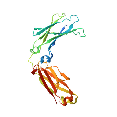
    8TTM, 8TUD

  • PubMed Abstract: 

    Redesigning protein-protein interfaces is an important tool for developing therapeutic strategies. Interfaces can be redesigned by in silico screening, which allows for efficient sampling of a large protein space before experimental validation. However, computational costs limit the number of combinations that can be reasonably sampled. Here, we present combinatorial tyrosine (Y)/serine (S) selection (combYSelect), a computational approach combining in silico determination of the change in binding free energy (ΔΔ G ) of an interface with a highly restricted library composed of just two amino acids, tyrosine and serine. We used combYSelect to design two immunoglobulin G (IgG) heterodimers-combYSelect1 (L368S/D399Y-K409S/T411Y) and combYSelect2 (D399Y/K447S-K409S/T411Y)-that exhibit near-optimal heterodimerization, without affecting IgG stability or function. We solved the crystal structures of these heterodimers and found that dynamic π-stacking interactions and polar contacts drive preferential heterodimeric interactions. Finally, we demonstrated the utility of our combYSelect heterodimers by engineering both a bispecific antibody and a cytokine trap for two unique therapeutic applications.


  • Organizational Affiliation
    • Department of Biochemistry, Emory University School of Medicine, Atlanta, GA 30322, USA.

Macromolecule Content 

  • Total Structure Weight: 55.22 kDa 
  • Atom Count: 3,545 
  • Modeled Residue Count: 414 
  • Deposited Residue Count: 464 
  • Unique protein chains: 2

Macromolecules

Find similar proteins by:|  3D Structure
Entity ID: 1
MoleculeChains  Sequence LengthOrganismDetailsImage
Immunoglobulin gamma-1 heavy chain232Homo sapiens x Mus musculus hybrid cell lineMutation(s): 2 
UniProt
Find proteins for P0DOX5 (Homo sapiens)
Explore P0DOX5 
Go to UniProtKB:  P0DOX5
Entity Groups
Sequence Clusters30% Identity50% Identity70% Identity90% Identity95% Identity100% Identity
UniProt GroupP0DOX5
Glycosylation
Glycosylation Sites: 1
Sequence Annotations
Expand
Reference Sequence
Find similar proteins by:|  3D Structure
Entity ID: 2
MoleculeChains  Sequence LengthOrganismDetailsImage
Immunoglobulin gamma-1 heavy chain232Homo sapiens x Mus musculus hybrid cell lineMutation(s): 2 
UniProt
Find proteins for P0DOX5 (Homo sapiens)
Explore P0DOX5 
Go to UniProtKB:  P0DOX5
Entity Groups
Sequence Clusters30% Identity50% Identity70% Identity90% Identity95% Identity100% Identity
UniProt GroupP0DOX5
Glycosylation
Glycosylation Sites: 1
Sequence Annotations
Expand
Reference Sequence

Oligosaccharides

Help  
Entity ID: 3
MoleculeChains Length2D Diagram GlycosylationD Interactions
2-acetamido-2-deoxy-beta-D-glucopyranose-(1-2)-alpha-D-mannopyranose-(1-3)-[2-acetamido-2-deoxy-beta-D-glucopyranose-(1-2)-alpha-D-mannopyranose-(1-6)]beta-D-mannopyranose-(1-4)-2-acetamido-2-deoxy-beta-D-glucopyranose-(1-4)-[alpha-L-fucopyranose-(1-6)]2-acetamido-2-deoxy-beta-D-glucopyranose
C, D
8N-Glycosylation
Glycosylation Resources
GlyTouCan: G80858MF
GlyCosmos: G80858MF
GlyGen: G80858MF

Experimental Data & Validation

Experimental Data

  • Method: X-RAY DIFFRACTION
  • Resolution: 2.51 Å
  • R-Value Free:  0.242 (Depositor), 0.246 (DCC) 
  • R-Value Work:  0.202 (Depositor), 0.212 (DCC) 
  • R-Value Observed: 0.206 (Depositor) 
Space Group: P 21 21 21
Unit Cell:
Length ( Å )Angle ( ˚ )
a = 49.782α = 90
b = 79.394β = 90
c = 142.902γ = 90
Software Package:
Software NamePurpose
PDB-REDOrefinement
PHENIXrefinement
PHENIXphasing
HKL-2000data scaling
HKL-2000data reduction

Structure Validation

View Full Validation Report


Entry History 

& Funding Information

Deposition Data


Funding OrganizationLocationGrant Number
National Institutes of Health/National Institute Of Allergy and Infectious Diseases (NIH/NIAID)United StatesR01AI149297

Revision History  (Full details and data files)

  • Version 1.0: 2024-06-26
    Type: Initial release
  • Version 1.1: 2024-10-30
    Changes: Structure summary