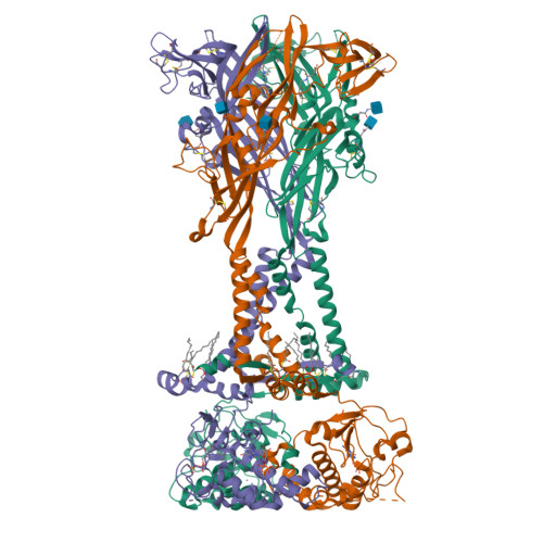
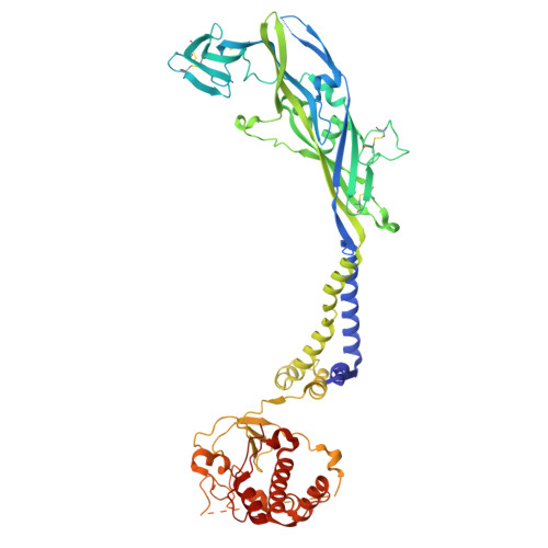
8TRA

Cryo-EM structure of the rat P2X7 receptor in complex with the allosteric antagonist GSK1482160


Experimental Data Snapshot

  • Method: ELECTRON MICROSCOPY
  • Resolution: 2.41 Å
  • Aggregation State: PARTICLE 
  • Reconstruction Method: SINGLE PARTICLE 

wwPDB Validation   3D Report Full Report


This is version 1.1 of the entry. See complete history


Literature

P2X 7 receptors exhibit at least three modes of allosteric antagonism.

Oken, A.C.Ditter, I.A.Lisi, N.E.Krishnamurthy, I.Godsey, M.H.Mansoor, S.E.

(2024) Sci Adv 10: eado5084-eado5084

  • DOI: https://doi.org/10.1126/sciadv.ado5084
  • Primary Citation of Related Structures:  
    8TR6, 8TR7, 8TR8, 8TRA, 8TRB, 8TRK

  • PubMed Abstract: 

    P2X receptors are trimeric ion channels activated by adenosine triphosphate (ATP) that contribute to pathophysiological processes ranging from asthma to neuropathic pain and neurodegeneration. A number of small-molecule antagonists have been identified for these important pharmaceutical targets. However, the molecular pharmacology of P2X receptors is poorly understood because of the chemically disparate nature of antagonists and their differential actions on the seven constituent subtypes. Here, we report high-resolution cryo-electron microscopy structures of the homomeric rat P2X 7 receptor bound to five previously known small-molecule allosteric antagonists and a sixth antagonist that we identify. Our structural, biophysical, and electrophysiological data define the molecular determinants of allosteric antagonism in this pharmacologically relevant receptor, revealing three distinct classes of antagonists that we call shallow, deep, and starfish. Starfish binders, exemplified by the previously unidentified antagonist methyl blue, represent a unique class of inhibitors with distinct functional properties that could be exploited to develop potent P2X 7 ligands with substantial clinical impact.


  • Organizational Affiliation

    Department of Chemical Physiology and Biochemistry, Oregon Health & Science University, Portland, OR 97239, USA.


Macromolecules
Find similar proteins by:  (by identity cutoff)  |  3D Structure
Entity ID: 1
MoleculeChains Sequence LengthOrganismDetailsImage
P2X purinoceptor 7
A, B, C
595RattusMutation(s): 0 
Gene Names: P2rx7
Membrane Entity: Yes 
UniProt
Find proteins for Q64663 (Rattus norvegicus)
Explore Q64663 
Go to UniProtKB:  Q64663
Entity Groups  
Sequence Clusters30% Identity50% Identity70% Identity90% Identity95% Identity100% Identity
UniProt GroupQ64663
Glycosylation
Glycosylation Sites: 2Go to GlyGen: Q64663-1
Sequence Annotations
Expand
  • Reference Sequence
Small Molecules
Ligands 6 Unique
IDChains Name / Formula / InChI Key2D Diagram3D Interactions
GDP (Subject of Investigation/LOI)
Query on GDP

Download Ideal Coordinates CCD File 
BA [auth C],
E [auth A],
Q [auth B]
GUANOSINE-5'-DIPHOSPHATE
C10 H15 N5 O11 P2
QGWNDRXFNXRZMB-UUOKFMHZSA-N
KF0 (Subject of Investigation/LOI)
Query on KF0

Download Ideal Coordinates CCD File 
AA [auth C],
D [auth A],
P [auth B]
N-{[2-chloro-3-(trifluoromethyl)phenyl]methyl}-1-methyl-5-oxo-L-prolinamide
C14 H14 Cl F3 N2 O2
BJEMSIVBBUBXMZ-JTQLQIEISA-N
PLM (Subject of Investigation/LOI)
Query on PLM

Download Ideal Coordinates CCD File 
GA [auth C]
HA [auth C]
IA [auth C]
J [auth A]
JA [auth C]
GA [auth C],
HA [auth C],
IA [auth C],
J [auth A],
JA [auth C],
K [auth A],
KA [auth C],
L [auth A],
M [auth A],
N [auth A],
V [auth B],
W [auth B],
X [auth B],
Y [auth B],
Z [auth B]
PALMITIC ACID
C16 H32 O2
IPCSVZSSVZVIGE-UHFFFAOYSA-N
NAG
Query on NAG

Download Ideal Coordinates CCD File 
EA [auth C]
FA [auth C]
H [auth A]
I [auth A]
T [auth B]
EA [auth C],
FA [auth C],
H [auth A],
I [auth A],
T [auth B],
U [auth B]
2-acetamido-2-deoxy-beta-D-glucopyranose
C8 H15 N O6
OVRNDRQMDRJTHS-FMDGEEDCSA-N
ZN (Subject of Investigation/LOI)
Query on ZN

Download Ideal Coordinates CCD File 
CA [auth C]
DA [auth C]
F [auth A]
G [auth A]
R [auth B]
CA [auth C],
DA [auth C],
F [auth A],
G [auth A],
R [auth B],
S [auth B]
ZINC ION
Zn
PTFCDOFLOPIGGS-UHFFFAOYSA-N
NA (Subject of Investigation/LOI)
Query on NA

Download Ideal Coordinates CCD File 
O [auth A]SODIUM ION
Na
FKNQFGJONOIPTF-UHFFFAOYSA-N
Experimental Data & Validation

Experimental Data

  • Method: ELECTRON MICROSCOPY
  • Resolution: 2.41 Å
  • Aggregation State: PARTICLE 
  • Reconstruction Method: SINGLE PARTICLE 

Structure Validation

View Full Validation Report


Entry History & Funding Information

Deposition Data


Funding OrganizationLocationGrant Number
National Institutes of Health/National Heart, Lung, and Blood Institute (NIH/NHLBI)United StatesR00HL138129
National Institutes of Health/National Institute of General Medical Sciences (NIH/NIGMS)United StatesDP2GM149551

Revision History  (Full details and data files)

  • Version 1.0: 2024-10-16
    Type: Initial release
  • Version 1.1: 2024-11-20
    Changes: Data collection