8TLN | pdb_00008tln

STRUCTURAL COMPARISON SUGGESTS THAT THERMOLYSIN AND RELATED NEUTRAL PROTEASES UNDERGO HINGE-BENDING MOTION DURING CATALYSIS


Experimental Data Snapshot

  • Method: X-RAY DIFFRACTION
  • Resolution: 1.60 Å
  • R-Value Work: 
    0.177 (DCC) 
  • R-Value Observed: 
    0.165 (Depositor) 

wwPDB Validation 3D Report Full Report

Validation slider image for 8TLN

This is version 1.4 of the entry. See complete history

Literature

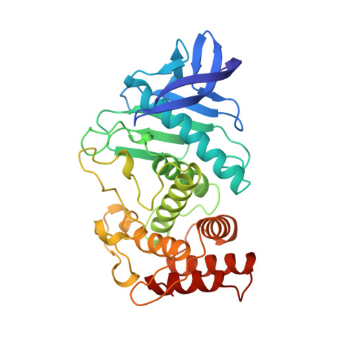
Structural comparison suggests that thermolysin and related neutral proteases undergo hinge-bending motion during catalysis.

Holland, D.R.Tronrud, D.E.Pley, H.W.Flaherty, K.M.Stark, W.Jansonius, J.N.McKay, D.B.Matthews, B.W.

(1992) Biochemistry 31: 11310-11316

  • DOI: https://doi.org/10.1021/bi00161a008
  • Primary Citation Related Structures: 
    8TLN

  • PubMed Abstract: 

    Crystal structures are known for three members of the bacterial neutral protease family: thermolysin from Bacillus thermoproteolyticus (TLN), the neutral protease from Bacillus cereus (NEU), and the elastase of Pseudomonas aeruginosa (PAE), both in free and ligand-bound forms. Each enzyme consists of an N-terminal and C-terminal domain with the active site formed at the junction of the two domains. Comparison of the different molecules reveals that the structure within each domain is well conserved, but there are substantial hinge-bending displacements (up to 16 degrees) of one domain relative to the other. These domain motions can be correlated with the presence or absence of bound inhibitor, as was previously observed in the specific example of PAE [Thayer, M.M., Flaherty, K.M., & McKay, D.B. (1991) J. Biol. Chem. 266, 2864-2871]. The binding of inhibitor appears to be associated with a reduction of the domain hinge-bending angle by 6-14 degrees and a closure of the "jaws" of the active site cleft by about 2 A. Crystallographic refinement of the structure of thermolysin suggests that electron density seen in the active site of the enzyme in the original structure determination probably corresponds to a bound dipeptide. Thus, the crystal structure appears to correspond to an enzyme-inhibitor or enzyme-product complex, rather than the free enzyme, as has previously been assumed.


  • Organizational Affiliation
    • Institute of Molecular Biology, University of Oregon, Eugene 97403.

Macromolecule Content 

  • Total Structure Weight: 34.93 kDa 
  • Atom Count: 2,621 
  • Modeled Residue Count: 316 
  • Deposited Residue Count: 316 
  • Unique protein chains: 1

Macromolecules

Find similar proteins by:|  3D Structure
Entity ID: 1
MoleculeChains  Sequence LengthOrganismDetailsImage
THERMOLYSINA [auth E]316Bacillus thermoproteolyticusMutation(s): 0 
EC: 3.4.24.27
UniProt
Find proteins for P00800 (Bacillus thermoproteolyticus)
Explore P00800 
Go to UniProtKB:  P00800
Entity Groups
Sequence Clusters30% Identity50% Identity70% Identity90% Identity95% Identity100% Identity
UniProt GroupP00800
Sequence Annotations
Expand
Reference Sequence

Small Molecules

Ligands 5 Unique
IDChains Name / Formula / InChI Key2D Diagram3D Interactions
LYS

Query on LYS


Download:Ideal Coordinates CCD File
C [auth E]LYSINE
C6 H15 N2 O2
KDXKERNSBIXSRK-YFKPBYRVSA-O
VAL

Query on VAL


Download:Ideal Coordinates CCD File
B [auth E]VALINE
C5 H11 N O2
KZSNJWFQEVHDMF-BYPYZUCNSA-N
DMS

Query on DMS


Download:Ideal Coordinates CCD File
I [auth E]DIMETHYL SULFOXIDE
C2 H6 O S
IAZDPXIOMUYVGZ-UHFFFAOYSA-N
ZN

Query on ZN


Download:Ideal Coordinates CCD File
H [auth E]ZINC ION
Zn
PTFCDOFLOPIGGS-UHFFFAOYSA-N
CA

Query on CA


Download:Ideal Coordinates CCD File
D [auth E],
E,
F [auth E],
G [auth E]
CALCIUM ION
Ca
BHPQYMZQTOCNFJ-UHFFFAOYSA-N

Experimental Data & Validation

Experimental Data

  • Method: X-RAY DIFFRACTION
  • Resolution: 1.60 Å
  • R-Value Work:  0.177 (DCC) 
  • R-Value Observed: 0.165 (Depositor) 
Space Group: P 61 2 2
Unit Cell:
Length ( Å )Angle ( ˚ )
a = 94.1α = 90
b = 94.1β = 90
c = 131.4γ = 120
Software Package:
Software NamePurpose
TNTrefinement

Structure Validation

View Full Validation Report


Entry History 

Deposition Data

Revision History  (Full details and data files)

  • Version 1.0: 1994-04-30
    Type: Initial release
  • Version 1.1: 2008-03-25
    Changes: Version format compliance
  • Version 1.2: 2011-07-13
    Changes: Version format compliance
  • Version 1.3: 2017-11-29
    Changes: Derived calculations, Other
  • Version 1.4: 2024-02-14
    Changes: Data collection, Database references, Derived calculations