8TJK

SAM-dependent methyltransferase RedM bound to SAH


Experimental Data Snapshot

  • Method: X-RAY DIFFRACTION
  • Resolution: 1.85 Å
  • R-Value Free: 0.229 
  • R-Value Work: 0.197 
  • R-Value Observed: 0.198 

wwPDB Validation   3D Report Full Report


Ligand Structure Quality Assessment 


This is version 1.1 of the entry. See complete history


Literature

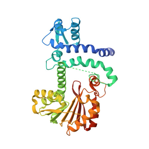
Structure of methyltransferase RedM that forms the dimethylpyrrolinium of the bisindole reductasporine.

Daniel-Ivad, P.Ryan, K.S.

(2023) J Biol Chem 300: 105520-105520

  • DOI: https://doi.org/10.1016/j.jbc.2023.105520
  • Primary Citation of Related Structures:  
    8TJI, 8TJJ, 8TJK

  • PubMed Abstract: 

    Bisindoles are biologically active natural products that arise from the oxidative dimerization of two molecules of l-tryptophan. In bacterial bisindole pathways, a core set of transformations is followed by the action of diverse tailoring enzymes that catalyze reactions that lead to diverse bisindole products. Among bisindoles, reductasporine is distinct due to its dimethylpyrrolinium structure. Its previously reported biosynthetic gene cluster encodes two unique tailoring enzymes, the imine reductase RedE and the dimethyltransferase RedM, which were shown to produce reductasporine from a common bisindole intermediate in recombinant E. coli. To gain more insight into the unique tailoring enzymes in reductasporine assembly, we reconstituted the biosynthetic pathway to reductasporine in vitro and then solved the 1.7 Å resolution structure of RedM. Our work reveals RedM adopts a variety of conformational changes with distinct open and closed conformations, and site-directed mutagenesis alongside sequence analysis identifies important active site residues. Finally, our work sets the stage for understanding how RedM evolved to react with a pyrrolinium scaffold and may enable the development of new dimethyltransferase catalysts.


  • Organizational Affiliation

    Department of Chemistry, The University of British Columbia, Vancouver, Canada.


Macromolecules
Find similar proteins by:  (by identity cutoff)  |  3D Structure
Entity ID: 1
MoleculeChains Sequence LengthOrganismDetailsImage
RedM
A, B
344uncultured bacteriumMutation(s): 0 
Gene Names: redM
UniProt
Find proteins for A0A0F7G196 (uncultured bacterium)
Explore A0A0F7G196 
Go to UniProtKB:  A0A0F7G196
Entity Groups  
Sequence Clusters30% Identity50% Identity70% Identity90% Identity95% Identity100% Identity
UniProt GroupA0A0F7G196
Sequence Annotations
Expand
  • Reference Sequence
Experimental Data & Validation

Experimental Data

  • Method: X-RAY DIFFRACTION
  • Resolution: 1.85 Å
  • R-Value Free: 0.229 
  • R-Value Work: 0.197 
  • R-Value Observed: 0.198 
  • Space Group: P 21 21 21
Unit Cell:
Length ( Å )Angle ( ˚ )
a = 61.833α = 90
b = 71.902β = 90
c = 144.841γ = 90
Software Package:
Software NamePurpose
PHENIXrefinement
Blu-Icedata collection
XDSdata reduction
Aimlessdata scaling
PHASERphasing
PHENIXmodel building

Structure Validation

View Full Validation Report


Ligand Structure Quality Assessment 


Entry History & Funding Information

Deposition Data


Funding OrganizationLocationGrant Number
Natural Sciences and Engineering Research Council (NSERC, Canada)CanadaRGPIN-2021-02626

Revision History  (Full details and data files)

  • Version 1.0: 2023-12-13
    Type: Initial release
  • Version 1.1: 2024-01-10
    Changes: Database references