8TH6

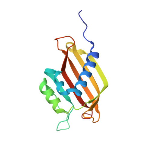
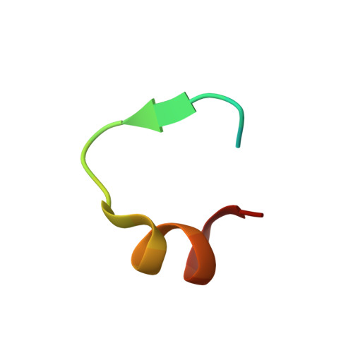
Crystal Structure of the G3BP1 NTF2-like domain bound to USP10 peptide


Experimental Data Snapshot

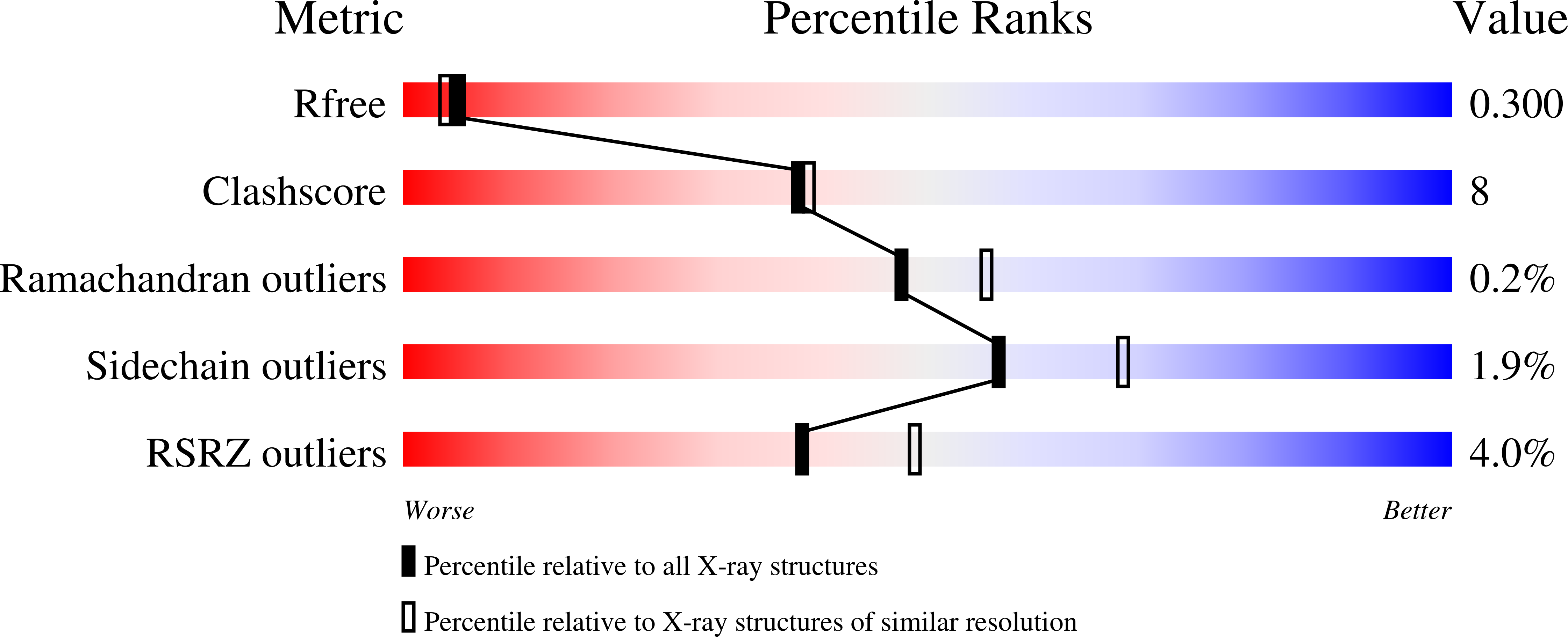
  • Method: X-RAY DIFFRACTION
  • Resolution: 2.34 Å
  • R-Value Free: 0.295 
  • R-Value Work: 0.213 
  • R-Value Observed: 0.217 

Starting Model: experimental
View more details

wwPDB Validation   3D Report Full Report


This is version 1.2 of the entry. See complete history


Literature

Interaction between host G3BP and viral nucleocapsid protein regulates SARS-CoV-2 replication and pathogenicity.

Yang, Z.Johnson, B.A.Meliopoulos, V.A.Ju, X.Zhang, P.Hughes, M.P.Wu, J.Koreski, K.P.Clary, J.E.Chang, T.C.Wu, G.Hixon, J.Duffner, J.Wong, K.Lemieux, R.Lokugamage, K.G.Alvarado, R.E.Crocquet-Valdes, P.A.Walker, D.H.Plante, K.S.Plante, J.A.Weaver, S.C.Kim, H.J.Meyers, R.Schultz-Cherry, S.Ding, Q.Menachery, V.D.Taylor, J.P.

(2024) Cell Rep 43: 113965-113965

  • DOI: https://doi.org/10.1016/j.celrep.2024.113965
  • Primary Citation of Related Structures:  
    8TH1, 8TH5, 8TH6, 8TH7

  • PubMed Abstract: 

    G3BP1/2 are paralogous proteins that promote stress granule formation in response to cellular stresses, including viral infection. The nucleocapsid (N) protein of severe acute respiratory syndrome coronavirus 2 (SARS-CoV-2) inhibits stress granule assembly and interacts with G3BP1/2 via an ITFG motif, including residue F17, in the N protein. Prior studies examining the impact of the G3PB1-N interaction on SARS-CoV-2 replication have produced inconsistent findings, and the role of this interaction in pathogenesis is unknown. Here, we use structural and biochemical analyses to define the residues required for G3BP1-N interaction and structure-guided mutagenesis to selectively disrupt this interaction. We find that N-F17A mutation causes highly specific loss of interaction with G3BP1/2. SARS-CoV-2 N-F17A fails to inhibit stress granule assembly in cells, has decreased viral replication, and causes decreased pathology in vivo. Further mechanistic studies indicate that the N-F17-mediated G3BP1-N interaction promotes infection by limiting sequestration of viral genomic RNA (gRNA) into stress granules.


  • Organizational Affiliation

    Department of Cell and Molecular Biology, St. Jude Children's Research Hospital, Memphis, TN, USA; Integrated Biomedical Sciences Program, University of Tennessee Health Science Center, Memphis, TN, USA.


Macromolecules
Find similar proteins by:  (by identity cutoff)  |  3D Structure
Entity ID: 1
MoleculeChains Sequence LengthOrganismDetailsImage
Ras GTPase-activating protein-binding protein 1
A, B, C, D
139Homo sapiensMutation(s): 0 
Gene Names: G3BP1G3BP
EC: 3.6.4.12 (PDB Primary Data), 3.6.4.13 (PDB Primary Data)
UniProt & NIH Common Fund Data Resources
Find proteins for Q13283 (Homo sapiens)
Explore Q13283 
Go to UniProtKB:  Q13283
PHAROS:  Q13283
GTEx:  ENSG00000145907 
Entity Groups  
Sequence Clusters30% Identity50% Identity70% Identity90% Identity95% Identity100% Identity
UniProt GroupQ13283
Sequence Annotations
Expand
  • Reference Sequence

Find similar proteins by:  Sequence   |   3D Structure  

Entity ID: 2
MoleculeChains Sequence LengthOrganismDetailsImage
Ubiquitin carboxyl-terminal hydrolase 10
E, F, G, H
24Homo sapiensMutation(s): 0 
Gene Names: USP10KIAA0190
EC: 3.4.19.12
UniProt & NIH Common Fund Data Resources
Find proteins for Q14694 (Homo sapiens)
Explore Q14694 
Go to UniProtKB:  Q14694
PHAROS:  Q14694
GTEx:  ENSG00000103194 
Entity Groups  
Sequence Clusters30% Identity50% Identity70% Identity90% Identity95% Identity100% Identity
UniProt GroupQ14694
Sequence Annotations
Expand
  • Reference Sequence
Small Molecules
Ligands 1 Unique
IDChains Name / Formula / InChI Key2D Diagram3D Interactions
EDO
Query on EDO

Download Ideal Coordinates CCD File 
I [auth A]
J [auth A]
K [auth A]
L [auth A]
M [auth A]
I [auth A],
J [auth A],
K [auth A],
L [auth A],
M [auth A],
N [auth A],
O [auth B],
P [auth B],
Q [auth B],
R [auth C],
S [auth D],
T [auth D],
U [auth D],
V [auth D],
W [auth H]
1,2-ETHANEDIOL
C2 H6 O2
LYCAIKOWRPUZTN-UHFFFAOYSA-N
Experimental Data & Validation

Experimental Data

  • Method: X-RAY DIFFRACTION
  • Resolution: 2.34 Å
  • R-Value Free: 0.295 
  • R-Value Work: 0.213 
  • R-Value Observed: 0.217 
  • Space Group: P 1 21 1
Unit Cell:
Length ( Å )Angle ( ˚ )
a = 58.39α = 90
b = 78.38β = 108.63
c = 73.41γ = 90
Software Package:
Software NamePurpose
REFMACrefinement
DIALSdata reduction
DIALSdata scaling
PHASERphasing

Structure Validation

View Full Validation Report


Entry History & Funding Information

Deposition Data


Funding OrganizationLocationGrant Number
Howard Hughes Medical Institute (HHMI)United StatesR35NS097974

Revision History  (Full details and data files)

  • Version 1.0: 2024-03-20
    Type: Initial release
  • Version 1.1: 2024-04-10
    Changes: Database references
  • Version 1.2: 2024-05-08
    Changes: Structure summary