Download Mendeley
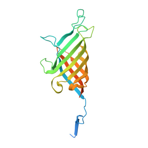
Dirigent isoflavene-forming PsPTS2: 3D structure, stereochemical, and kinetic characterization comparison with pterocarpan-forming PsPTS1 homolog in pea.
Meng, Q., Moinuddin, S.G.A., Celoy, R.M., Smith, C.A., Young, R.P., Costa, M.A., Freeman, R.A., Fukaya, M., Kim, D.N., Cort, J.R., Hawes, M.C., van Etten, H.D., Pandey, P., Chittiboyina, A.G., Ferreira, D., Davin, L.B., Lewis, N.G.(2024) J Biol Chem 300: 105647-105647
- PubMed: 38219818 Search on PubMedSearch on PubMed Central
- DOI: https://doi.org/10.1016/j.jbc.2024.105647
- Primary Citation Related Structures:
8TH2 - PubMed Abstract:
Pea phytoalexins (-)-maackiain and (+)-pisatin have opposite C6a/C11a configurations, but biosynthetically how this occurs is unknown. Pea dirigent-protein (DP) PsPTS2 generates 7,2'-dihydroxy-4',5'-methylenedioxyisoflav-3-ene (DMDIF), and stereoselectivity toward four possible 7,2'-dihydroxy-4',5'-methylenedioxyisoflavan-4-ol (DMDI) stereoisomers was investigated. Stereoisomer configurations were determined using NMR spectroscopy, electronic circular dichroism, and molecular orbital analyses. PsPTS2 efficiently converted cis-(3R,4R)-DMDI into DMDIF 20-fold faster than the trans-(3R,4S)-isomer. The 4R-configured substrate's near β-axial OH orientation significantly enhanced its leaving group abilities in generating A-ring mono-quinone methide (QM), whereas 4S-isomer's α-equatorial-OH was a poorer leaving group. Docking simulations indicated that the 4R-configured β-axial OH was closest to Asp 51 , whereas 4S-isomer's α-equatorial OH was further away. Neither cis-(3S,4S)- nor trans-(3S,4R)-DMDIs were substrates, even with the former having C3/C4 stereochemistry as in (+)-pisatin. PsPTS2 used cis-(3R,4R)-7,2'-dihydroxy-4'-methoxyisoflavan-4-ol [cis-(3R,4R)-DMI] and C3/C4 stereoisomers to give 2',7-dihydroxy-4'-methoxyisoflav-3-ene (DMIF). DP homologs may exist in licorice (Glycyrrhiza pallidiflora) and tree legume Bolusanthus speciosus, as DMIF occurs in both species. PsPTS1 utilized cis-(3R,4R)-DMDI to give (-)-maackiain 2200-fold more efficiently than with cis-(3R,4R)-DMI to give (-)-medicarpin. PsPTS1 also slowly converted trans-(3S,4R)-DMDI into (+)-maackiain, reflecting the better 4R configured OH leaving group. PsPTS2 and PsPTS1 provisionally provide the means to enable differing C6a and C11a configurations in (+)-pisatin and (-)-maackiain, via identical DP-engendered mono-QM bound intermediate generation, which PsPTS2 either re-aromatizes to give DMDIF or PsPTS1 intramolecularly cyclizes to afford (-)-maackiain. Substrate docking simulations using PsPTS2 and PsPTS1 indicate cis-(3R,4R)-DMDI binds in the anti-configuration in PsPTS2 to afford DMDIF, and the syn-configuration in PsPTS1 to give maackiain.
- Institute of Biological Chemistry, Washington State University, Pullman, Washington, USA.
Organizational Affiliation: