An alternative broad-specificity pathway for glycan breakdown in bacteria.
Nasseri, S.A., Lazarski, A.C., Lemmer, I.L., Zhang, C.Y., Brencher, E., Chen, H.M., Sim, L., Panwar, D., Betschart, L., Worrall, L.J., Brumer, H., Strynadka, N.C.J., Withers, S.G.(2024) Nature 631: 199-206
- PubMed: 38898276 Search on PubMedSearch on PubMed Central
- DOI: https://doi.org/10.1038/s41586-024-07574-y
- Primary Citation Related Structures:
8TCD, 8TCR, 8TCS, 8TCT, 8TDA, 8TDE, 8TDF, 8TDH, 8TDI, 8V31 - PubMed Abstract:
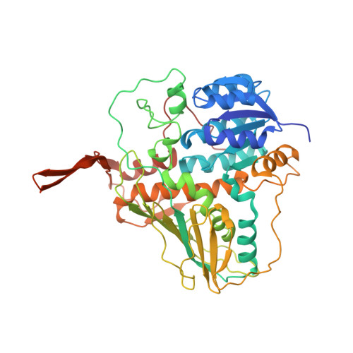
The vast majority of glycosidases characterized to date follow one of the variations of the 'Koshland' mechanisms 1 to hydrolyse glycosidic bonds through substitution reactions. Here we describe a large-scale screen of a human gut microbiome metagenomic library using an assay that selectively identifies non-Koshland glycosidase activities 2 . Using this, we identify a cluster of enzymes with extremely broad substrate specificities and thoroughly characterize these, mechanistically and structurally. These enzymes not only break glycosidic linkages of both α and β stereochemistry and multiple connectivities, but also cleave substrates that are not hydrolysed by standard glycosidases. These include thioglycosides, such as the glucosinolates from plants, and pseudoglycosidic bonds of pharmaceuticals such as acarbose. This is achieved through a distinct mechanism of hydrolysis that involves oxidation/reduction and elimination/hydration steps, each catalysed by enzyme modules that are in many cases interchangeable between organisms and substrate classes. Homologues of these enzymes occur in both Gram-positive and Gram-negative bacteria associated with the gut microbiome and other body parts, as well as other environments, such as soil and sea. Such alternative step-wise mechanisms appear to constitute largely unrecognized but abundant pathways for glycan degradation as part of the metabolism of carbohydrates in bacteria.
- Department of Chemistry, University of British Columbia, Vancouver, British Columbia, Canada.
Organizational Affiliation: