8TAO

Quis and CDPPB bound active mGlu5


Experimental Data Snapshot

  • Method: ELECTRON MICROSCOPY
  • Resolution: 2.90 Å
  • Aggregation State: PARTICLE 
  • Reconstruction Method: SINGLE PARTICLE 

wwPDB Validation   3D Report Full Report


This is version 1.2 of the entry. See complete history


Literature

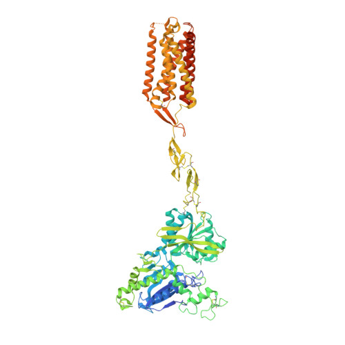
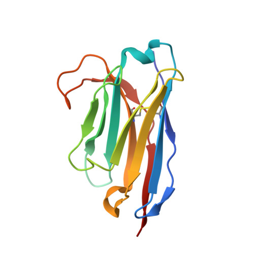
Stepwise activation of a metabotropic glutamate receptor.

Krishna Kumar, K.Wang, H.Habrian, C.Latorraca, N.R.Xu, J.O'Brien, E.S.Zhang, C.Montabana, E.Koehl, A.Marqusee, S.Isacoff, E.Y.Kobilka, B.K.

(2024) Nature 629: 951-956

  • DOI: https://doi.org/10.1038/s41586-024-07327-x
  • Primary Citation of Related Structures:  
    8T6J, 8T7H, 8T8M, 8TAO

  • PubMed Abstract: 

    Metabotropic glutamate receptors belong to a family of G protein-coupled receptors that are obligate dimers and possess a large extracellular ligand-binding domain that is linked via a cysteine-rich domain to their 7-transmembrane domain 1 . Upon activation, these receptors undergo a large conformational change to transmit the ligand binding signal from the extracellular ligand-binding domain to the G protein-coupling 7-transmembrane domain 2 . In this manuscript, we propose a model for a sequential, multistep activation mechanism of metabotropic glutamate receptor subtype 5. We present a series of structures in lipid nanodiscs, from inactive to fully active, including agonist-bound intermediate states. Further, using bulk and single-molecule fluorescence imaging, we reveal distinct receptor conformations upon allosteric modulator and G protein binding.


  • Organizational Affiliation

    Department of Molecular and Cellular Physiology, Stanford University School of Medicine, Stanford, CA, USA. kaavyak@stanford.edu.


Macromolecules
Find similar proteins by:  (by identity cutoff)  |  3D Structure
Entity ID: 1
MoleculeChains Sequence LengthOrganismDetailsImage
Metabotropic glutamate receptor 5
A, B
881Homo sapiensMutation(s): 0 
Gene Names: GRM5GPRC1EMGLUR5
Membrane Entity: Yes 
UniProt & NIH Common Fund Data Resources
Find proteins for P41594 (Homo sapiens)
Explore P41594 
Go to UniProtKB:  P41594
PHAROS:  P41594
GTEx:  ENSG00000168959 
Entity Groups  
Sequence Clusters30% Identity50% Identity70% Identity90% Identity95% Identity100% Identity
UniProt GroupP41594
Sequence Annotations
Expand
  • Reference Sequence
Find similar proteins by:  (by identity cutoff)  |  3D Structure
Entity ID: 2
MoleculeChains Sequence LengthOrganismDetailsImage
Nb43
C, D
123Lama glamaMutation(s): 0 
Entity Groups  
Sequence Clusters30% Identity50% Identity70% Identity90% Identity95% Identity100% Identity
Sequence Annotations
Expand
  • Reference Sequence
Small Molecules
Experimental Data & Validation

Experimental Data

  • Method: ELECTRON MICROSCOPY
  • Resolution: 2.90 Å
  • Aggregation State: PARTICLE 
  • Reconstruction Method: SINGLE PARTICLE 

Structure Validation

View Full Validation Report


Entry History & Funding Information

Deposition Data


Funding OrganizationLocationGrant Number
National Institutes of Health/National Institute of Neurological Disorders and Stroke (NIH/NINDS)United StatesNS028471

Revision History  (Full details and data files)

  • Version 1.0: 2023-10-11
    Type: Initial release
  • Version 1.1: 2024-05-29
    Changes: Database references
  • Version 1.2: 2024-06-05
    Changes: Database references