8T6L | pdb_00008t6l

Cryo-EM structure of rat cardiac sodium channel NaV1.5 with batrachotoxin analog BTX-B


Experimental Data Snapshot

  • Method: ELECTRON MICROSCOPY
  • Resolution: 3.30 Å
  • Aggregation State: PARTICLE 
  • Reconstruction Method: SINGLE PARTICLE 

Starting Model: experimental
View more details

wwPDB Validation 3D Report Full Report

Validation slider image for 8T6L

This is version 1.2 of the entry. See complete history

Literature

Dual receptor-sites reveal the structural basis for hyperactivation of sodium channels by poison-dart toxin batrachotoxin.

Tonggu, L.Wisedchaisri, G.Gamal El-Din, T.M.Lenaeus, M.J.Logan, M.M.Toma, T.Du Bois, J.Zheng, N.Catterall, W.A.

(2024) Nat Commun 15: 2306-2306

  • DOI: https://doi.org/10.1038/s41467-024-45958-w
  • Primary Citation Related Structures: 
    8T6L

  • PubMed Abstract: 

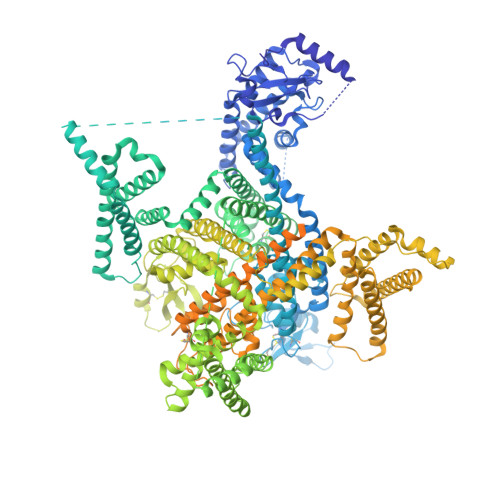
    The poison dart toxin batrachotoxin is exceptional for its high potency and toxicity, and for its multifaceted modification of the function of voltage-gated sodium channels. By using cryogenic electron microscopy, we identify two homologous, but nonidentical receptor sites that simultaneously bind two molecules of toxin, one at the interface between Domains I and IV, and the other at the interface between Domains III and IV of the cardiac sodium channel. Together, these two bound toxin molecules stabilize α/π helical conformation in the S6 segments that gate the pore, and one of the bound BTX-B molecules interacts with the crucial Lys1421 residue that is essential for sodium conductance and selectivity via an apparent water-bridged hydrogen bond. Overall, our structure provides insight into batrachotoxin's potency, efficacy, and multifaceted functional effects on voltage-gated sodium channels via a dual receptor site mechanism.


  • Organizational Affiliation
    • Department of Pharmacology, University of Washington, Seattle, WA, 98195, USA.

Macromolecule Content 

  • Total Structure Weight: 231.78 kDa 
  • Atom Count: 10,988 
  • Modeled Residue Count: 1,237 
  • Deposited Residue Count: 1,874 
  • Unique protein chains: 1

Macromolecules

Find similar proteins by:|  3D Structure
Entity ID: 1
MoleculeChains  Sequence LengthOrganismDetailsImage
Sodium channel protein type 5 subunit alpha,Green fluorescent protein1,874Rattus norvegicusAequorea victoria
This entity is chimeric
Mutation(s): 2 
Gene Names: Scn5aGFP
Membrane Entity: Yes 
UniProt
Find proteins for P15389 (Rattus norvegicus)
Explore P15389 
Go to UniProtKB:  P15389
Find proteins for P42212 (Aequorea victoria)
Explore P42212 
Go to UniProtKB:  P42212
Entity Groups
Sequence Clusters30% Identity50% Identity70% Identity90% Identity95% Identity100% Identity
UniProt GroupsP42212P15389
Glycosylation
Glycosylation Sites: 7Go to GlyGen: P15389-1
Sequence Annotations
Expand
Reference Sequence

Oligosaccharides

Help  
Entity ID: 2
MoleculeChains Length2D Diagram GlycosylationD Interactions
alpha-D-mannopyranose-(1-3)-beta-D-mannopyranose-(1-4)-2-acetamido-2-deoxy-beta-D-glucopyranose-(1-3)-2-acetamido-2-deoxy-beta-D-glucopyranose
B
4N-Glycosylation
Entity ID: 3
MoleculeChains Length2D Diagram GlycosylationD Interactions
2-acetamido-2-deoxy-beta-D-glucopyranose-(1-4)-2-acetamido-2-deoxy-beta-D-glucopyranose
C, D
2N-Glycosylation
Glycosylation Resources
GlyTouCan: G42666HT
GlyCosmos: G42666HT
GlyGen: G42666HT
Entity ID: 4
MoleculeChains Length2D Diagram GlycosylationD Interactions
alpha-D-mannopyranose-(1-6)-alpha-D-mannopyranose-(1-6)-beta-D-mannopyranose-(1-4)-2-acetamido-2-deoxy-beta-D-glucopyranose-(1-4)-2-acetamido-2-deoxy-beta-D-glucopyranose
E
5N-Glycosylation
Glycosylation Resources
GlyTouCan: G94626GC
GlyCosmos: G94626GC
GlyGen: G94626GC

Small Molecules

Ligands 5 Unique
IDChains Name / Formula / InChI Key2D Diagram3D Interactions
LBN

Query on LBN


Download:Ideal Coordinates CCD File
I [auth A]
J [auth A]
K [auth A]
L [auth A]
M [auth A]
I [auth A],
J [auth A],
K [auth A],
L [auth A],
M [auth A],
N [auth A],
O [auth A],
P [auth A],
Q [auth A],
R [auth A]
1-palmitoyl-2-oleoyl-sn-glycero-3-phosphocholine
C42 H82 N O8 P
WTJKGGKOPKCXLL-VYOBOKEXSA-N
9Z9

Query on 9Z9


Download:Ideal Coordinates CCD File
DA [auth A],
EA [auth A]
(3beta,14beta,17beta,25R)-3-[4-methoxy-3-(methoxymethyl)butoxy]spirost-5-en
C34 H56 O5
CEEBZAXXSRFQIC-GZSGZGDASA-N
YIJ
(Subject of Investigation/LOI)

Query on YIJ


Download:Ideal Coordinates CCD File
FA [auth A],
GA [auth A]
(1R)-1-[(5aR,7aR,9R,11aS,11bS,12R,13aR)-9,12-dihydroxy-2,11a-dimethyl-1,2,3,4,7a,8,9,10,11,11a,12,13-dodecahydro-7H-9,11b-epoxy-13a,5a-prop[1]enophenanthro[2,1-f][1,4]oxazepin-14-yl]ethyl benzoate
C31 H39 N O6
BQDUFJGPBNLKPK-UDTYFQFOSA-N
Y01

Query on Y01


Download:Ideal Coordinates CCD File
AA [auth A]
BA [auth A]
CA [auth A]
S [auth A]
T [auth A]
AA [auth A],
BA [auth A],
CA [auth A],
S [auth A],
T [auth A],
U [auth A],
V [auth A],
W [auth A],
X [auth A],
Y [auth A],
Z [auth A]
CHOLESTEROL HEMISUCCINATE
C31 H50 O4
WLNARFZDISHUGS-MIXBDBMTSA-N
NAG

Query on NAG


Download:Ideal Coordinates CCD File
F [auth A],
G [auth A],
H [auth A]
2-acetamido-2-deoxy-beta-D-glucopyranose
C8 H15 N O6
OVRNDRQMDRJTHS-FMDGEEDCSA-N

Experimental Data & Validation

Experimental Data

  • Method: ELECTRON MICROSCOPY
  • Resolution: 3.30 Å
  • Aggregation State: PARTICLE 
  • Reconstruction Method: SINGLE PARTICLE 
EM Software:
TaskSoftware PackageVersion
MODEL REFINEMENTPHENIX
RECONSTRUCTIONcryoSPARC3.3.1

Structure Validation

View Full Validation Report


Entry History 

& Funding Information

Deposition Data


Funding OrganizationLocationGrant Number
National Institutes of Health/National Heart, Lung, and Blood Institute (NIH/NHLBI)United StatesR01 HL112808
National Institutes of Health/National Institute of Neurological Disorders and Stroke (NIH/NINDS)United StatesR35 NS111573
National Institutes of Health/National Institute of General Medical Sciences (NIH/NIGMS)United StatesR01 GM117263
Howard Hughes Medical Institute (HHMI)United States--

Revision History  (Full details and data files)

  • Version 1.0: 2024-03-06
    Type: Initial release
  • Version 1.1: 2024-03-27
    Changes: Database references
  • Version 1.2: 2024-11-20
    Changes: Data collection, Structure summary