


2.07 Angstrom CryoEM Structure of Heavy Chain Apoferritin from Mus Musculus From 200kV Microscope
Liu, Y., Pintilie, G., Chiu, W.To be published.
Experimental Data Snapshot
Starting Model: experimental
View more details
wwPDB Validation 3D Report Full Report

Entity ID: 1 | |||||
|---|---|---|---|---|---|
| Molecule | Chains | Sequence Length | Organism | Details | Image |
| Ferritin heavy chain, N-terminally processed | 172 | Mus musculus | Mutation(s): 0 Gene Names: Fth1, Fth EC: 1.16.3.1 | 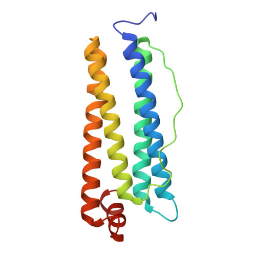 | |
UniProt & NIH Common Fund Data Resources | |||||
IMPC: MGI:95588 | |||||
Entity Groups | |||||
| Sequence Clusters | 30% Identity50% Identity70% Identity90% Identity95% Identity100% Identity | ||||
| UniProt Group | P09528 | ||||
Sequence AnnotationsExpand | |||||
Reference Sequence | |||||
| Ligands 1 Unique | |||||
|---|---|---|---|---|---|
| ID | Chains | Name / Formula / InChI Key | 2D Diagram | 3D Interactions | |
| FE (Subject of Investigation/LOI) Download:Ideal Coordinates CCD File | AA [auth C] BA [auth D] CA [auth E] DA [auth F] EA [auth G] | FE (III) ION Fe VTLYFUHAOXGGBS-UHFFFAOYSA-N |  | ||
| Task | Software Package | Version |
|---|---|---|
| MODEL REFINEMENT | UCSF ChimeraX | 1.6 |
| Funding Organization | Location | Grant Number |
|---|---|---|
| National Institutes of Health/National Institute of General Medical Sciences (NIH/NIGMS) | United States | U24GM129541 |


