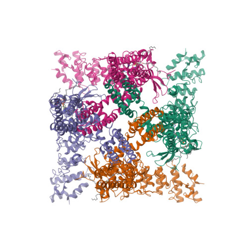
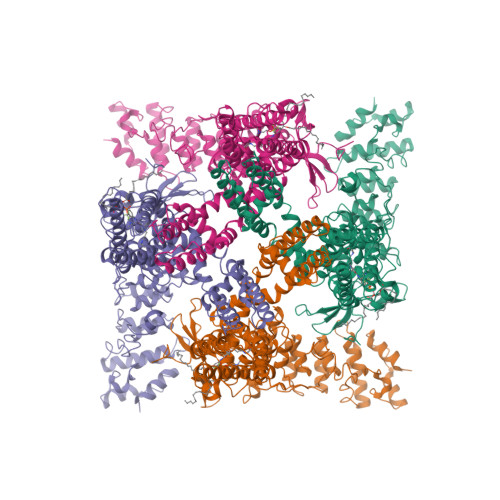
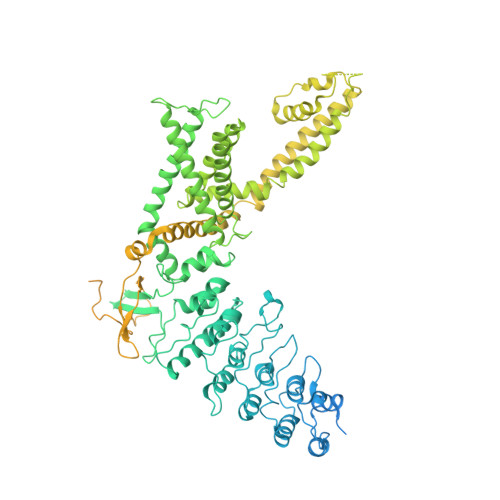
8T1F

Cryo-EM structure of full-length human TRPV4 in complex with antagonist HC-067047

  • Classification: MEMBRANE PROTEIN
  • Organism(s): Homo sapiens, Human betaherpesvirus 5
  • Expression System: Homo sapiens
  • Mutation(s): No 
  • Membrane Protein: Yes  PDBTMMemProtMDmpstruc

  • Deposited: 2023-06-02 Released: 2023-07-05 
  • Deposition Author(s): Talyzina, I.A., Nadezhdin, K.D., Neuberger, A., Sobolevsky, A.I.
  • Funding Organization(s): National Institutes of Health/National Institute of Arthritis and Musculoskeletal and Skin Diseases (NIH/NIAMS), National Institutes of Health/National Cancer Institute (NIH/NCI), National Institutes of Health/National Institute of Neurological Disorders and Stroke (NIH/NINDS), German Research Foundation (DFG), National Institutes of Health/National Heart, Lung, and Blood Institute (NIH/NHLBI)

Experimental Data Snapshot

  • Method: ELECTRON MICROSCOPY
  • Resolution: 3.49 Å
  • Aggregation State: PARTICLE 
  • Reconstruction Method: SINGLE PARTICLE 

wwPDB Validation   3D Report Full Report


This is version 1.1 of the entry. See complete history


Literature

Structure of human TRPV4 in complex with GTPase RhoA.

Nadezhdin, K.D.Talyzina, I.A.Parthasarathy, A.Neuberger, A.Zhang, D.X.Sobolevsky, A.I.

(2023) Nat Commun 14: 3733-3733

  • DOI: https://doi.org/10.1038/s41467-023-39346-z
  • Primary Citation of Related Structures:  
    8T1B, 8T1C, 8T1D, 8T1E, 8T1F

  • PubMed Abstract: 

    Transient receptor potential (TRP) channel TRPV4 is a polymodal cellular sensor that responds to moderate heat, cell swelling, shear stress, and small-molecule ligands. It is involved in thermogenesis, regulation of vascular tone, bone homeostasis, renal and pulmonary functions. TRPV4 is implicated in neuromuscular and skeletal disorders, pulmonary edema, and cancers, and represents an important drug target. The cytoskeletal remodeling GTPase RhoA has been shown to suppress TRPV4 activity. Here, we present a structure of the human TRPV4-RhoA complex that shows RhoA interaction with the membrane-facing surface of the TRPV4 ankyrin repeat domains. The contact interface reveals residues that are mutated in neuropathies, providing an insight into the disease pathogenesis. We also identify the binding sites of the TRPV4 agonist 4α-PDD and the inhibitor HC-067047 at the base of the S1-S4 bundle, and show that agonist binding leads to pore opening, while channel inhibition involves a π-to-α transition in the pore-forming helix S6. Our structures elucidate the interaction interface between hTRPV4 and RhoA, as well as residues at this interface that are involved in TRPV4 disease-causing mutations. They shed light on TRPV4 activation and inhibition and provide a template for the design of future therapeutics for treatment of TRPV4-related diseases.


  • Organizational Affiliation

    Department of Biochemistry and Molecular Biophysics, Columbia University, New York, NY, 10032, USA.


Macromolecules
Find similar proteins by:  (by identity cutoff)  |  3D Structure
Entity ID: 1
MoleculeChains Sequence LengthOrganismDetailsImage
Transient receptor potential cation channel subfamily V member 4/Enhanced green fluorescent protein chimera
A, B, C, D
1,132Homo sapiensHuman betaherpesvirus 5
This entity is chimeric
Mutation(s): 0 
Gene Names: TRPV4VRL2VROACegfp
Membrane Entity: Yes 
UniProt & NIH Common Fund Data Resources
Find proteins for Q9HBA0 (Homo sapiens)
Explore Q9HBA0 
Go to UniProtKB:  Q9HBA0
PHAROS:  Q9HBA0
GTEx:  ENSG00000111199 
Entity Groups  
Sequence Clusters30% Identity50% Identity70% Identity90% Identity95% Identity100% Identity
UniProt GroupQ9HBA0
Sequence Annotations
Expand
  • Reference Sequence
Small Molecules
Ligands 2 Unique
IDChains Name / Formula / InChI Key2D Diagram3D Interactions
POV
Query on POV

Download Ideal Coordinates CCD File 
F [auth A],
H [auth B],
J [auth C],
L [auth D]
(2S)-3-(hexadecanoyloxy)-2-[(9Z)-octadec-9-enoyloxy]propyl 2-(trimethylammonio)ethyl phosphate
C42 H82 N O8 P
WTJKGGKOPKCXLL-PFDVCBLKSA-N
X7N (Subject of Investigation/LOI)
Query on X7N

Download Ideal Coordinates CCD File 
E [auth A],
G [auth B],
I [auth C],
K [auth D]
2-methyl-1-[3-(morpholin-4-yl)propyl]-5-phenyl-N-[3-(trifluoromethyl)phenyl]-1H-pyrrole-3-carboxamide
C26 H28 F3 N3 O2
NCZYSQOTAYFTNM-UHFFFAOYSA-N
Binding Affinity Annotations 
IDSourceBinding Affinity
X7N BindingDB:  8T1F IC50: min: 17, max: 180 (nM) from 7 assay(s)
Experimental Data & Validation

Experimental Data

  • Method: ELECTRON MICROSCOPY
  • Resolution: 3.49 Å
  • Aggregation State: PARTICLE 
  • Reconstruction Method: SINGLE PARTICLE 
EM Software:
TaskSoftware PackageVersion
MODEL REFINEMENTPHENIX1.18

Structure Validation

View Full Validation Report


Entry History & Funding Information

Deposition Data


Funding OrganizationLocationGrant Number
National Institutes of Health/National Institute of Arthritis and Musculoskeletal and Skin Diseases (NIH/NIAMS)United StatesR01 AR078814
National Institutes of Health/National Cancer Institute (NIH/NCI)United StatesR01 CA206573
National Institutes of Health/National Institute of Neurological Disorders and Stroke (NIH/NINDS)United StatesR01 NS083660
National Institutes of Health/National Institute of Neurological Disorders and Stroke (NIH/NINDS)United StatesR01 NS107253
German Research Foundation (DFG)Germany464295817
National Institutes of Health/National Heart, Lung, and Blood Institute (NIH/NHLBI)United StatesR01 HL096647

Revision History  (Full details and data files)

  • Version 1.0: 2023-07-05
    Type: Initial release
  • Version 1.1: 2024-06-19
    Changes: Data collection