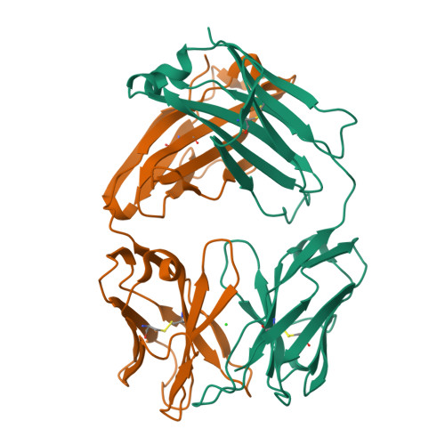
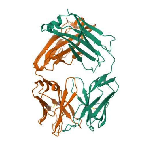
8T0O

Fab from mAb RB2AT_87


Experimental Data Snapshot

  • Method: X-RAY DIFFRACTION
  • Resolution: 2.30 Å
  • R-Value Free: 0.257 
  • R-Value Work: 0.215 
  • R-Value Observed: 0.219 

Starting Model: experimental
View more details

wwPDB Validation   3D Report Full Report


This is version 1.2 of the entry. See complete history


Literature

Generation and Study of Antibodies against Two Triangular Trimers Derived from A beta.

Kreutzer, A.G.Malonis, R.J.Parrocha, C.M.T.Tong, K.Guaglianone, G.Nguyen, J.T.Diab, M.N.Lai, J.R.Nowick, J.S.

(2024) Pept Sci (Hoboken) 116

  • DOI: https://doi.org/10.1002/pep2.24333
  • Primary Citation of Related Structures:  
    8T0O

  • PubMed Abstract: 

    Monoclonal antibodies (mAbs) that target the P-amyloid peptide (Aβ) are important Alzheimer's disease research tools and are now being used as Alzheimer's disease therapies. Conformation-specific mAbs that target oligomeric and fibrillar Aβ assemblies are of particular interest, as these assemblies are associated with Alzheimer's disease pathogenesis and progression. This paper reports the generation of rabbit mAbs against two different triangular trimers derived from Aβ. These antibodies are the first mAbs generated against Aβ oligomer mimics in which the high-resolution structures of the oligomers are known. We describe the isolation of the mAbs using single B-cell sorting of peripheral blood mononuclear cells (PBMCs) from immunized rabbits, the selectivity of the mAbs for the triangular trimers, the immunoreactivity of the mAbs with aggregated Aβ 42 , and the immunoreactivity of the mAbs in brain tissue from the 5xFAD Alzheimer's disease mouse model. The characterization of these mAbs against structurally defined trimers derived from Aβ enhances understanding of antibody-amyloid recognition and may benefit the development of diagnostics and immunotherapies in Alzheimer's disease.


  • Organizational Affiliation

    Department of Chemistry, University of California Irvine, Irvine, CA 92697.


Macromolecules
Find similar proteins by:  (by identity cutoff)  |  3D Structure
Entity ID: 1
MoleculeChains Sequence LengthOrganismDetailsImage
RB2AT_87 Fab Light chainA [auth L]217Oryctolagus cuniculusMutation(s): 0 
Entity Groups  
Sequence Clusters30% Identity50% Identity70% Identity90% Identity95% Identity100% Identity
Sequence Annotations
Expand
  • Reference Sequence
Find similar proteins by:  (by identity cutoff)  |  3D Structure
Entity ID: 2
MoleculeChains Sequence LengthOrganismDetailsImage
RB2AT_87 Fab Heavy chainB [auth H]217Oryctolagus cuniculusMutation(s): 0 
Entity Groups  
Sequence Clusters30% Identity50% Identity70% Identity90% Identity95% Identity100% Identity
Sequence Annotations
Expand
  • Reference Sequence
Small Molecules
Ligands 1 Unique
IDChains Name / Formula / InChI Key2D Diagram3D Interactions
CL
Query on CL

Download Ideal Coordinates CCD File 
C [auth H]CHLORIDE ION
Cl
VEXZGXHMUGYJMC-UHFFFAOYSA-M
Experimental Data & Validation

Experimental Data

  • Method: X-RAY DIFFRACTION
  • Resolution: 2.30 Å
  • R-Value Free: 0.257 
  • R-Value Work: 0.215 
  • R-Value Observed: 0.219 
  • Space Group: P 31 2 1
Unit Cell:
Length ( Å )Angle ( ˚ )
a = 111.358α = 90
b = 111.358β = 90
c = 79.156γ = 120
Software Package:
Software NamePurpose
PHENIXrefinement
XDSdata reduction
Aimlessdata scaling
PHASERphasing

Structure Validation

View Full Validation Report


Entry History & Funding Information

Deposition Data


Funding OrganizationLocationGrant Number
National Institutes of Health/National Institute on Aging (NIH/NIA)United StatesAG062296

Revision History  (Full details and data files)

  • Version 1.0: 2024-03-27
    Type: Initial release
  • Version 1.1: 2024-05-01
    Changes: Database references
  • Version 1.2: 2024-10-23
    Changes: Structure summary