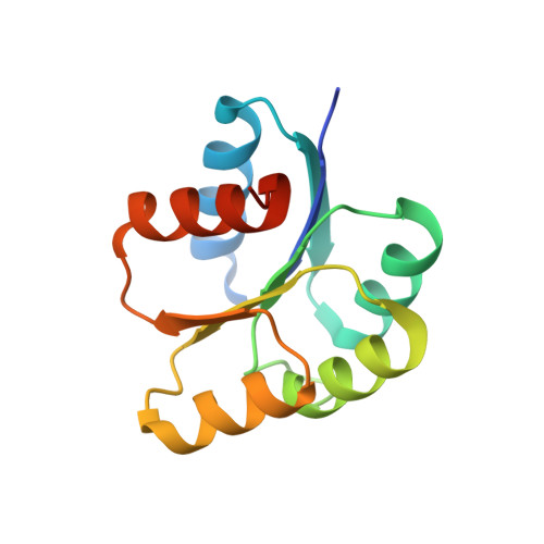
Insights into DNA-binding motifs and mechanisms of Francisella tularensis novicida two-component system response regulator proteins QseB, KdpE, and BfpR.
Gaddy, K.E., Bensch, E.M., Cavanagh, J., Milton, M.E.(2024) Biochem Biophys Res Commun 722: 150150-150150
- PubMed: 38805787
- DOI: https://doi.org/10.1016/j.bbrc.2024.150150
- Primary Citation of Related Structures:
8SVZ - PubMed Abstract:
Two component system bacterial response regulators are typically DNA-binding proteins which enable the genetic regulation of many adaptive bacterial behaviors. Despite structural similarity across response regulator families, there is a diverse array of DNA-binding mechanisms. Bacteria usually encode several dozen two-component system response regulators, but Francisella tularensis only encodes three. Due to their simplified response regulatory network, Francisella species are a model for studying the role of response regulator proteins in virulence. Here, we show that Francisella response regulators QseB, KdpE, and BfpR all utilize different DNA-binding mechanisms. Our evidence suggests that QseB follows a simple mechanism whereby it binds a single inverted repeat sequence with a higher affinity upon phosphorylation. This behavior is independent of whether QseB is a positive or negative regulator of the gene as demonstrated by qseB and priM promoter sequences, respectively. Similarly, KdpE binds DNA more tightly upon phosphorylation, but also exhibits a cooperative binding isotherm. While we propose a KdpE binding site, it is possible that KdpE has a complex DNA-binding mechanism potentially involving multiple copies of KdpE being recruited to a promoter region. Finally, we show that BfpR appears to bind a region of its own promoter sequence with a lower affinity upon phosphorylation. Further structural and enzymatic work will need to be performed to deconvolute the KdpE and BfpR binding mechanisms.
Organizational Affiliation:
Department of Biochemistry and Molecular Biology, Brody School of Medicine, East Carolina University, Greenville, NC, USA.


