8SST

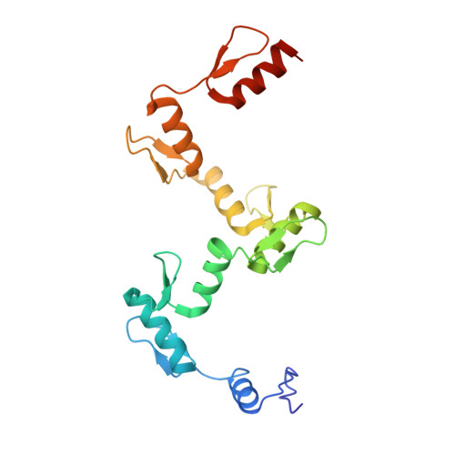
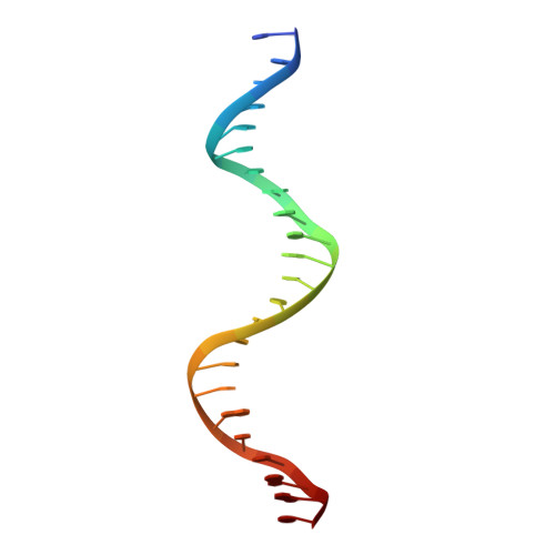
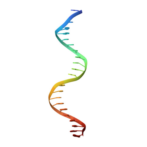
ZnFs 1-7 of CCCTC-binding factor (CTCF) K365T Mutant Complexed with 23mer


Experimental Data Snapshot

  • Method: X-RAY DIFFRACTION
  • Resolution: 2.19 Å
  • R-Value Free: 0.243 
  • R-Value Work: 0.220 
  • R-Value Observed: 0.221 

Starting Model: experimental
View more details

wwPDB Validation   3D Report Full Report


This is version 1.1 of the entry. See complete history


Literature

Structures of CTCF-DNA complexes including all 11 zinc fingers.

Yang, J.Horton, J.R.Liu, B.Corces, V.G.Blumenthal, R.M.Zhang, X.Cheng, X.

(2023) Nucleic Acids Res 51: 8447-8462

  • DOI: https://doi.org/10.1093/nar/gkad594
  • Primary Citation of Related Structures:  
    8SSQ, 8SSR, 8SSS, 8SST, 8SSU

  • PubMed Abstract: 

    The CCCTC-binding factor (CTCF) binds tens of thousands of enhancers and promoters on mammalian chromosomes by means of its 11 tandem zinc finger (ZF) DNA-binding domain. In addition to the 12-15-bp CORE sequence, some of the CTCF binding sites contain 5' upstream and/or 3' downstream motifs. Here, we describe two structures for overlapping portions of human CTCF, respectively, including ZF1-ZF7 and ZF3-ZF11 in complex with DNA that incorporates the CORE sequence together with either 3' downstream or 5' upstream motifs. Like conventional tandem ZF array proteins, ZF1-ZF7 follow the right-handed twist of the DNA, with each finger occupying and recognizing one triplet of three base pairs in the DNA major groove. ZF8 plays a unique role, acting as a spacer across the DNA minor groove and positioning ZF9-ZF11 to make cross-strand contacts with DNA. We ascribe the difference between the two subgroups of ZF1-ZF7 and ZF8-ZF11 to residues at the two positions -6 and -5 within each finger, with small residues for ZF1-ZF7 and bulkier and polar/charged residues for ZF8-ZF11. ZF8 is also uniquely rich in basic amino acids, which allows salt bridges to DNA phosphates in the minor groove. Highly specific arginine-guanine and glutamine-adenine interactions, used to recognize G:C or A:T base pairs at conventional base-interacting positions of ZFs, also apply to the cross-strand interactions adopted by ZF9-ZF11. The differences between ZF1-ZF7 and ZF8-ZF11 can be rationalized structurally and may contribute to recognition of high-affinity CTCF binding sites.


  • Organizational Affiliation

    Department of Epigenetics and Molecular Carcinogenesis, University of Texas MD Anderson Cancer Center, Houston, TX 77030, USA.


Macromolecules

Find similar proteins by:  (by identity cutoff)  |  3D Structure
Entity ID: 1
MoleculeChains Sequence LengthOrganismDetailsImage
Transcriptional repressor CTCF
A, D
203Homo sapiensMutation(s): 1 
Gene Names: CTCF
UniProt & NIH Common Fund Data Resources
Find proteins for P49711 (Homo sapiens)
Explore P49711 
Go to UniProtKB:  P49711
PHAROS:  P49711
GTEx:  ENSG00000102974 
Entity Groups  
Sequence Clusters30% Identity50% Identity70% Identity90% Identity95% Identity100% Identity
UniProt GroupP49711
Sequence Annotations
Expand
  • Reference Sequence

Find similar nucleic acids by:  Sequence   |   3D Structure  

Entity ID: 2
MoleculeChains LengthOrganismImage
DNA Strand (23mer) I
B, E
23Homo sapiens
Sequence Annotations
Expand
  • Reference Sequence

Find similar nucleic acids by:  Sequence   |   3D Structure  

Entity ID: 3
MoleculeChains LengthOrganismImage
DNA Strand (23mer) II
C, F
23Homo sapiens
Sequence Annotations
Expand
  • Reference Sequence
Small Molecules
Ligands 2 Unique
IDChains Name / Formula / InChI Key2D Diagram3D Interactions
ZN
Query on ZN

Download Ideal Coordinates CCD File 
G [auth A]
H [auth A]
I [auth A]
J [auth A]
K [auth A]
G [auth A],
H [auth A],
I [auth A],
J [auth A],
K [auth A],
L [auth A],
M [auth A],
Q [auth D],
R [auth D],
S [auth D],
T [auth D],
U [auth D],
V [auth D],
W [auth D]
ZINC ION
Zn
PTFCDOFLOPIGGS-UHFFFAOYSA-N
EDO
Query on EDO

Download Ideal Coordinates CCD File 
N [auth B],
O [auth B],
P [auth C],
X [auth E]
1,2-ETHANEDIOL
C2 H6 O2
LYCAIKOWRPUZTN-UHFFFAOYSA-N
Experimental Data & Validation

Experimental Data

  • Method: X-RAY DIFFRACTION
  • Resolution: 2.19 Å
  • R-Value Free: 0.243 
  • R-Value Work: 0.220 
  • R-Value Observed: 0.221 
  • Space Group: C 1 2 1
Unit Cell:
Length ( Å )Angle ( ˚ )
a = 160.561α = 90
b = 41.596β = 105.61
c = 135.215γ = 90
Software Package:
Software NamePurpose
SERGUIdata collection
PHENIXrefinement
HKL-2000data reduction
HKL-2000data scaling
PHENIXphasing

Structure Validation

View Full Validation Report


Entry History & Funding Information

Deposition Data


Funding OrganizationLocationGrant Number
National Institutes of Health/National Institute of General Medical Sciences (NIH/NIGMS)United StatesGM049245-23
Cancer Prevention and Research Institute of Texas (CPRIT)United StatesRR160029

Revision History  (Full details and data files)

  • Version 1.0: 2023-08-02
    Type: Initial release
  • Version 1.1: 2023-09-20
    Changes: Data collection, Database references