8SNP | pdb_00008snp

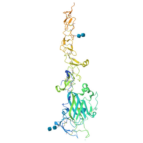
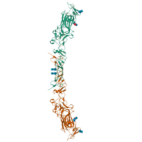
Crystal structure of mouse Netrin-1 in complex with samarium ions


Experimental Data Snapshot

  • Method: X-RAY DIFFRACTION
  • Resolution: 3.40 Å
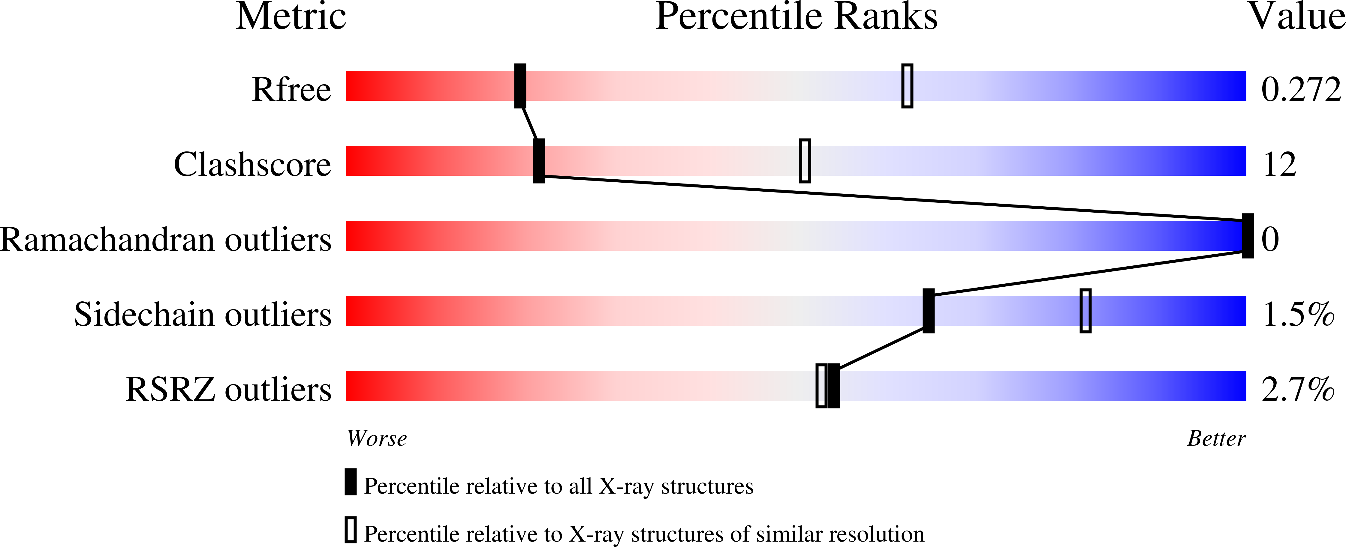
  • R-Value Free: 
    0.268 (Depositor), 0.270 (DCC) 
  • R-Value Work: 
    0.250 (Depositor), 0.250 (DCC) 
  • R-Value Observed: 
    0.251 (Depositor) 

Starting Model: experimental
View more details

wwPDB Validation   3D Report Full Report


This is version 1.2 of the entry. See complete history


Literature

Identifying the molecular basis of Laminin N-terminal domain Ca 2+ binding using a hybrid approach.

Legare, S.Heide, F.Gabir, H.Rafiei, F.Meier, M.Padilla-Meier, G.P.Koch, M.Stetefeld, J.

(2024) Biophys J 123: 2422-2430

  • DOI: https://doi.org/10.1016/j.bpj.2024.06.005
  • Primary Citation of Related Structures:  
    8SNP

  • PubMed Abstract: 

    Ca 2+ is a highly abundant ion involved in numerous biological processes, particularly in multicellular eukaryotic organisms where it exerts many of these functions through interactions with Ca 2+ binding proteins. The laminin N-terminal (LN) domain is found in members of the laminin and netrin protein families where it plays a critical role in the function of these proteins. The LN domain of laminins and netrins is a Ca 2+ binding domain and in many cases requires Ca 2+ to perform its biological function. Here, we conduct a detailed examination of the molecular basis of the LN domain Ca 2+ interaction combining structural, computational, bioinformatics, and biophysical techniques. By combining computational and bioinformatic techniques with x-ray crystallography we explore the molecular basis of the LN domain Ca 2+ interaction and identify a conserved sequence present in Ca 2+ binding LN domains. These findings enable a sequence-based prediction of LN domain Ca 2+ binding ability. We use thermal shift assays and isothermal titration calorimetry to explore the biophysical properties of the LN domain Ca 2+ interaction. We show that the netrin-1 LN domain exhibits a high affinity and specificity for Ca 2+ , which structurally stabilizes the LN domain. This study elucidates the molecular foundation of the LN domain Ca 2+ binding interaction and provides a detailed functional characterization of this essential interaction, advancing our understanding of protein-Ca 2+ dynamics within the context of the LN domain.


  • Organizational Affiliation

    Department of Chemistry, University of Manitoba, Winnipeg, Manitoba, Canada. Electronic address: legares@myumanitoba.ca.


Macromolecules
Find similar proteins by:  (by identity cutoff)  |  3D Structure
Entity ID: 1
MoleculeChains Sequence LengthOrganismDetailsImage
Netrin-1604Mus musculusMutation(s): 0 
Gene Names: Ntn1
UniProt
Find proteins for O09118 (Mus musculus)
Explore O09118 
Go to UniProtKB:  O09118
Entity Groups  
Sequence Clusters30% Identity50% Identity70% Identity90% Identity95% Identity100% Identity
UniProt GroupO09118
Glycosylation
Glycosylation Sites: 3Go to GlyGen: O09118-1
Sequence Annotations
Expand
  • Reference Sequence
Oligosaccharides

Help

Entity ID: 2
MoleculeChains Length2D Diagram Glycosylation3D Interactions
2-acetamido-2-deoxy-beta-D-glucopyranose-(1-4)-[alpha-L-fucopyranose-(1-6)]2-acetamido-2-deoxy-beta-D-glucopyranoseB [auth D]3N-Glycosylation
Glycosylation Resources
GlyTouCan:  G21290RB
GlyCosmos:  G21290RB
GlyGen:  G21290RB
Entity ID: 3
MoleculeChains Length2D Diagram Glycosylation3D Interactions
2-acetamido-2-deoxy-beta-D-glucopyranose-(1-4)-2-acetamido-2-deoxy-beta-D-glucopyranose
C
2N-Glycosylation
Glycosylation Resources
GlyTouCan:  G42666HT
GlyCosmos:  G42666HT
GlyGen:  G42666HT
Experimental Data & Validation

Experimental Data

  • Method: X-RAY DIFFRACTION
  • Resolution: 3.40 Å
  • R-Value Free:  0.268 (Depositor), 0.270 (DCC) 
  • R-Value Work:  0.250 (Depositor), 0.250 (DCC) 
  • R-Value Observed: 0.251 (Depositor) 
Space Group: P 32 2 1
Unit Cell:
Length ( Å )Angle ( ˚ )
a = 69.5α = 90
b = 69.5β = 90
c = 333.641γ = 120
Software Package:
Software NamePurpose
PHENIXrefinement
XDSdata reduction
XSCALEdata scaling
PHASERphasing

Structure Validation

View Full Validation Report


Entry History & Funding Information

Deposition Data


Funding OrganizationLocationGrant Number
Canadian Institutes of Health Research (CIHR)Canada--

Revision History  (Full details and data files)

  • Version 1.0: 2024-02-07
    Type: Initial release
  • Version 1.1: 2024-10-23
    Changes: Structure summary
  • Version 1.2: 2025-03-19
    Changes: Database references