8SMU | pdb_00008smu

Integral fusion of the HtaA CR2 domain from Corynebacterium diphtheriae within EGFP


Experimental Data Snapshot

  • Method: X-RAY DIFFRACTION
  • Resolution: 2.45 Å
  • R-Value Free: 
    0.233 (Depositor), 0.223 (DCC) 
  • R-Value Work: 
    0.209 (Depositor), 0.205 (DCC) 
  • R-Value Observed: 
    0.210 (Depositor) 

Starting Models: experimental
View more details

wwPDB Validation 3D Report Full Report

Validation slider image for 8SMU

Ligand Structure Quality Assessment 


This is version 2.0 of the entry. See complete history

Literature

Development and atomic structure of a new fluorescence-based sensor to probe heme transfer in bacterial pathogens.

Mahoney, B.J.Goring, A.K.Wang, Y.Dasika, P.Zhou, A.Grossbard, E.Cascio, D.Loo, J.A.Clubb, R.T.

(2023) J Inorg Biochem 249: 112368-112368

  • DOI: https://doi.org/10.1016/j.jinorgbio.2023.112368
  • Primary Citation Related Structures: 
    8SMU

  • PubMed Abstract: 

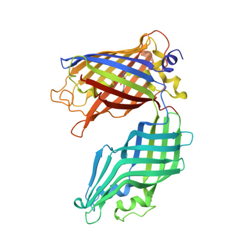
    Heme is the most abundant source of iron in the human body and is actively scavenged by bacterial pathogens during infections. Corynebacterium diphtheriae and other species of actinobacteria scavenge heme using cell wall associated and secreted proteins that contain Conserved Region (CR) domains. Here we report the development of a fluorescent sensor to measure heme transfer from the C-terminal CR domain within the HtaA protein (CR2) to other hemoproteins within the heme-uptake system. The sensor contains the CR2 domain inserted into the β2 to β3 turn of the Enhanced Green Fluorescent Protein (EGFP). A 2.45 Å crystal structure reveals the basis of heme binding to the CR2 domain via iron-tyrosyl coordination and shares conserved structural features with CR domains present in Corynebacterium glutamicum. The structure and small angle X-ray scattering experiments are consistent with the sensor adopting a V-shaped structure that exhibits only small fluctuations in inter-domain positioning. We demonstrate heme transfer from the sensor to the CR domains located within the HtaA or HtaB proteins in the heme-uptake system as measured by a ∼ 60% increase in sensor fluorescence and native mass spectrometry.


  • Organizational Affiliation
    • Department of Chemistry and Biochemistry, University of California, Los Angeles, 611 Charles E. Young Drive East, Los Angeles, CA 90095, USA; UCLA-DOE Institute of Genomics and Proteomics, University of California, Los Angeles, 611 Charles E. Young Drive East, Los Angeles, CA 90095, USA.

Macromolecule Content 

  • Total Structure Weight: 181.67 kDa 
  • Atom Count: 12,400 
  • Modeled Residue Count: 1,576 
  • Deposited Residue Count: 1,612 
  • Unique protein chains: 1

Macromolecules

Find similar proteins by:|  3D Structure
Entity ID: 1
MoleculeChains  Sequence LengthOrganismDetailsImage
HtaACR2 integral fusion within enhanced green fluorescent protein
A, B, C, D
403Aequorea victoriaCorynebacterium diphtheriae NCTC 13129Mutation(s): 1 
Gene Names: GFPDIP0625
UniProt
Find proteins for P42212 (Aequorea victoria)
Explore P42212 
Go to UniProtKB:  P42212
Find proteins for Q6NIZ1 (Corynebacterium diphtheriae (strain ATCC 700971 / NCTC 13129 / Biotype gravis))
Explore Q6NIZ1 
Go to UniProtKB:  Q6NIZ1
Entity Groups
Sequence Clusters30% Identity50% Identity70% Identity90% Identity95% Identity100% Identity
UniProt GroupsP42212Q6NIZ1
Sequence Annotations
Expand
Reference Sequence

Small Molecules

Ligands 4 Unique
IDChains Name / Formula / InChI Key2D Diagram3D Interactions
HEM
(Subject of Investigation/LOI)

Query on HEM


Download:Ideal Coordinates CCD File
E [auth A],
FA [auth D],
O [auth B],
V [auth C]
PROTOPORPHYRIN IX CONTAINING FE
C34 H32 Fe N4 O4
KABFMIBPWCXCRK-RGGAHWMASA-L
1PE

Query on 1PE


Download:Ideal Coordinates CCD File
IA [auth D],
K [auth A],
S [auth B],
Z [auth C]
PENTAETHYLENE GLYCOL
C10 H22 O6
JLFNLZLINWHATN-UHFFFAOYSA-N
GOL

Query on GOL


Download:Ideal Coordinates CCD File
AA [auth C]
BA [auth C]
CA [auth C]
DA [auth C]
F [auth A]
AA [auth C],
BA [auth C],
CA [auth C],
DA [auth C],
F [auth A],
G [auth A],
GA [auth D],
H [auth A],
HA [auth D],
I [auth A],
J [auth A],
JA [auth D],
KA [auth D],
L [auth A],
LA [auth D],
M [auth A],
P [auth B],
Q [auth B],
R [auth B],
T [auth B],
W [auth C],
X [auth C],
Y [auth C]
GLYCEROL
C3 H8 O3
PEDCQBHIVMGVHV-UHFFFAOYSA-N
CL

Query on CL


Download:Ideal Coordinates CCD File
EA [auth C],
MA [auth D],
N [auth A],
U [auth B]
CHLORIDE ION
Cl
VEXZGXHMUGYJMC-UHFFFAOYSA-M
Modified Residues  1 Unique
IDChains TypeFormula2D DiagramParent
CRO
Query on CRO
A, B, C, D
L-PEPTIDE LINKINGC15 H17 N3 O5THR, TYR, GLY

Experimental Data & Validation

Experimental Data

  • Method: X-RAY DIFFRACTION
  • Resolution: 2.45 Å
  • R-Value Free:  0.233 (Depositor), 0.223 (DCC) 
  • R-Value Work:  0.209 (Depositor), 0.205 (DCC) 
  • R-Value Observed: 0.210 (Depositor) 
Space Group: P 1 21 1
Unit Cell:
Length ( Å )Angle ( ˚ )
a = 63.826α = 90
b = 272.783β = 114.22
c = 73.518γ = 90
Software Package:
Software NamePurpose
autoPROCdata processing
pointlessdata scaling
Aimlessdata scaling
STARANISOdata scaling
BUSTERrefinement
XDSdata reduction
PHASERphasing

Structure Validation

View Full Validation Report


Ligand Structure Quality Assessment 


Entry History 

& Funding Information

Deposition Data


Funding OrganizationLocationGrant Number
National Institutes of Health/National Institute Of Allergy and Infectious Diseases (NIH/NIAID)United StatesR01-AI161828

Revision History  (Full details and data files)

  • Version 1.0: 2023-09-27
    Type: Initial release
  • Version 1.1: 2023-10-04
    Changes: Database references
  • Version 1.2: 2023-11-15
    Changes: Data collection
  • Version 1.3: 2024-11-13
    Changes: Structure summary
  • Version 2.0: 2026-03-18
    Changes: Polymer sequence