8SLC | pdb_00008slc

High Affinity nanobodies against GFP


Experimental Data Snapshot

  • Method: X-RAY DIFFRACTION
  • Resolution: 2.97 Å
  • R-Value Free: 
    0.241 (Depositor), 0.239 (DCC) 
  • R-Value Work: 
    0.220 (Depositor), 0.218 (DCC) 
  • R-Value Observed: 
    0.221 (Depositor) 

Starting Models: experimental
View more details

wwPDB Validation 3D Report Full Report

Validation slider image for 8SLC

This is version 2.0 of the entry. See complete history

Literature

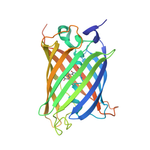
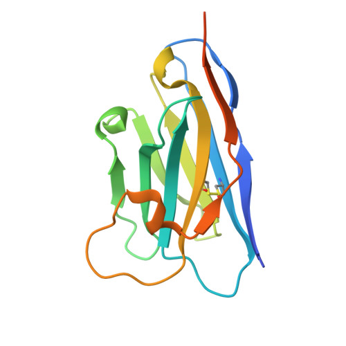
Unique mechanisms to increase structural stability and enhance antigen binding in nanobodies.

Ketaren, N.E.Fridy, P.C.Malashkevich, V.Sanyal, T.Brillantes, M.Thompson, M.K.Oren, D.A.Bonanno, J.B.Sali, A.Almo, S.C.Chait, B.T.Rout, M.P.

(2025) Structure 33: 677-690.e5

  • DOI: https://doi.org/10.1016/j.str.2025.01.019
  • Primary Citation Related Structures: 
    8G0I, 8SFS, 8SFV, 8SFX, 8SFZ, 8SG3, 8SLC

  • PubMed Abstract: 

    Nanobodies are single domain antibody variants proving themselves to be compelling tools for research, disease diagnostics, and as therapeutics targeting a myriad of disease agents. However, despite this potential, their mechanisms of paratope presentation and structural stabilization have not been fully explored. Here, we show that unlike monoclonal antibodies, a nanobody repertoire maximizes sampling of an antigen surface by binding a single antigen in at least three different orientations, which are correlated with their paratope composition. Structure-guided reengineering of several nanobodies reveals that a single point mutation within the paratope or a highly conserved region of a nanobody's framework 3 (FR3) can markedly improve antigen affinity, nanobody stability, or both. Conversely, we show the negative impact on antigen affinity when "over-stabilizing" nanobodies. Collectively our results provide a universal strategy to tune a nanobody's affinity by modifying specific residues that can readily be applied to guide nanobody optimization and functionalization.


  • Organizational Affiliation
    • Laboratory of Cellular and Structural Biology, The Rockefeller University, New York, NY 10065, USA. Electronic address: nketaren@rockefeller.edu.

Macromolecule Content 

  • Total Structure Weight: 90.49 kDa 
  • Atom Count: 5,638 
  • Modeled Residue Count: 712 
  • Deposited Residue Count: 802 
  • Unique protein chains: 2

Macromolecules

Find similar proteins by:|  3D Structure
Entity ID: 1
MoleculeChains  Sequence LengthOrganismDetailsImage
Green fluorescent protein
A, B
253Aequorea victoriaMutation(s): 1 
Gene Names: GFP
UniProt
Find proteins for P42212 (Aequorea victoria)
Explore P42212 
Go to UniProtKB:  P42212
Entity Groups
Sequence Clusters30% Identity50% Identity70% Identity90% Identity95% Identity100% Identity
UniProt GroupP42212
Sequence Annotations
Expand
Reference Sequence
Find similar proteins by:|  3D Structure
Entity ID: 2
MoleculeChains  Sequence LengthOrganismDetailsImage
LaG43
C, D
148Lama glamaMutation(s): 0 

Small Molecules

Ligands 3 Unique
IDChains Name / Formula / InChI Key2D Diagram3D Interactions
GOL

Query on GOL


Download:Ideal Coordinates CCD File
E [auth A],
H [auth B],
I [auth B],
J [auth C]
GLYCEROL
C3 H8 O3
PEDCQBHIVMGVHV-UHFFFAOYSA-N
CL

Query on CL


Download:Ideal Coordinates CCD File
F [auth A],
K [auth C],
L [auth C],
M [auth D]
CHLORIDE ION
Cl
VEXZGXHMUGYJMC-UHFFFAOYSA-M
NA

Query on NA


Download:Ideal Coordinates CCD File
G [auth A]SODIUM ION
Na
FKNQFGJONOIPTF-UHFFFAOYSA-N
Modified Residues  1 Unique
IDChains TypeFormula2D DiagramParent
CR2
Query on CR2
A, B
L-PEPTIDE LINKINGC13 H13 N3 O4GLY, TYR, GLY

Experimental Data & Validation

Experimental Data

  • Method: X-RAY DIFFRACTION
  • Resolution: 2.97 Å
  • R-Value Free:  0.241 (Depositor), 0.239 (DCC) 
  • R-Value Work:  0.220 (Depositor), 0.218 (DCC) 
  • R-Value Observed: 0.221 (Depositor) 
Space Group: P 63
Unit Cell:
Length ( Å )Angle ( ˚ )
a = 149.435α = 90
b = 149.435β = 90
c = 127.013γ = 120
Software Package:
Software NamePurpose
PHENIXrefinement
SCALEPACKdata scaling
DENZOdata reduction
PHASERphasing

Structure Validation

View Full Validation Report


Entry History 

& Funding Information

Deposition Data


Funding OrganizationLocationGrant Number
National Institutes of Health/National Institute of General Medical Sciences (NIH/NIGMS)United States--

Revision History  (Full details and data files)

  • Version 1.0: 2024-05-22
    Type: Initial release
  • Version 1.1: 2024-11-06
    Changes: Structure summary
  • Version 1.2: 2025-06-04
    Changes: Database references
  • Version 2.0: 2026-03-18
    Changes: Polymer sequence