8SJI | pdb_00008sji

Apo Structure of Computationally Designed Homotrimer TP1


Experimental Data Snapshot

  • Method: X-RAY DIFFRACTION
  • Resolution: 1.50 Å
  • R-Value Free: 
    0.206 (Depositor), 0.209 (DCC) 
  • R-Value Work: 
    0.168 (Depositor), 0.171 (DCC) 
  • R-Value Observed: 
    0.170 (Depositor) 

Starting Model: experimental
View more details

wwPDB Validation 3D Report Full Report

Validation slider image for 8SJI

This is version 1.2 of the entry. See complete history

Literature

Atomically Accurate Design of Metalloproteins with Predefined Coordination Geometries.

Hoffnagle, A.M.Tezcan, F.A.

(2023) J Am Chem Soc 145: 14208-14214

  • DOI: https://doi.org/10.1021/jacs.3c04047
  • Primary Citation Related Structures: 
    8SJF, 8SJG, 8SJH, 8SJI

  • PubMed Abstract: 

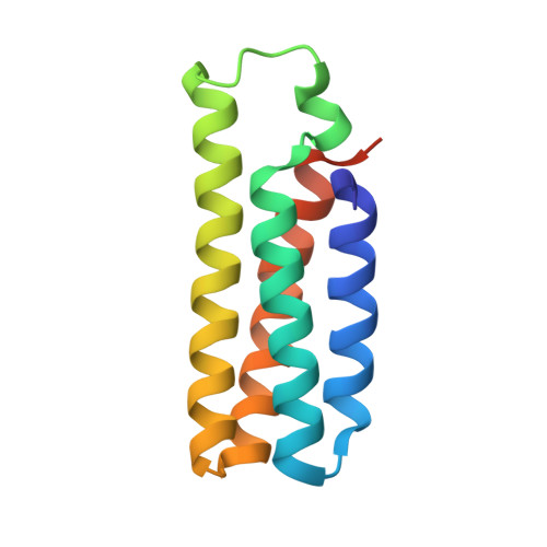
    We report a new computational protein design method for the construction of oligomeric protein assemblies around metal centers with predefined coordination geometries. We apply this method to design two homotrimeric assemblies, Tet4 and TP1, with tetrahedral and trigonal-pyramidal tris(histidine) metal coordination geometries, respectively, and demonstrate that both assemblies form the targeted metal centers with ≤0.2 Å accuracy. Although Tet4 and TP1 are constructed from the same parent protein building block, they are distinct in terms of their overall architectures, the environment surrounding the metal centers, and their metal-based reactivities, illustrating the versatility of our approach.


  • Organizational Affiliation
    • Department of Chemistry and Biochemistry, University of California, San Diego, La Jolla, California 92093, United States.

Macromolecule Content 

  • Total Structure Weight: 37.67 kDa 
  • Atom Count: 2,891 
  • Modeled Residue Count: 329 
  • Deposited Residue Count: 342 
  • Unique protein chains: 1

Macromolecules

Find similar proteins by:|  3D Structure
Entity ID: 1
MoleculeChains  Sequence LengthOrganismDetailsImage
Topoisomerase 1
A, B, C
114synthetic constructMutation(s): 0 

Experimental Data & Validation

Experimental Data

  • Method: X-RAY DIFFRACTION
  • Resolution: 1.50 Å
  • R-Value Free:  0.206 (Depositor), 0.209 (DCC) 
  • R-Value Work:  0.168 (Depositor), 0.171 (DCC) 
  • R-Value Observed: 0.170 (Depositor) 
Space Group: C 1 2 1
Unit Cell:
Length ( Å )Angle ( ˚ )
a = 95.06α = 90
b = 77.43β = 106.584
c = 47.44γ = 90
Software Package:
Software NamePurpose
PHENIXrefinement
XDSdata reduction
XDSdata scaling
PHASERphasing

Structure Validation

View Full Validation Report


Entry History 

& Funding Information

Deposition Data


Funding OrganizationLocationGrant Number
National Institutes of Health/National Institute of General Medical Sciences (NIH/NIGMS)United StatesR01-GM138884

Revision History  (Full details and data files)

  • Version 1.0: 2023-07-05
    Type: Initial release
  • Version 1.1: 2023-07-12
    Changes: Database references
  • Version 1.2: 2024-05-22
    Changes: Data collection