8SJ9 | pdb_00008sj9

Crystal structure of the H1 hemagglutinin COBRA X6


Experimental Data Snapshot

  • Method: X-RAY DIFFRACTION
  • Resolution: 3.25 Å
  • R-Value Free: 
    0.276 (Depositor), 0.277 (DCC) 
  • R-Value Work: 
    0.221 (Depositor), 0.221 (DCC) 
  • R-Value Observed: 
    0.224 (Depositor) 

Starting Model: in silico
View more details

wwPDB Validation 3D Report Full Report

Validation slider image for 8SJ9

Ligand Structure Quality Assessment 


This is version 1.4 of the entry. See complete history

Literature

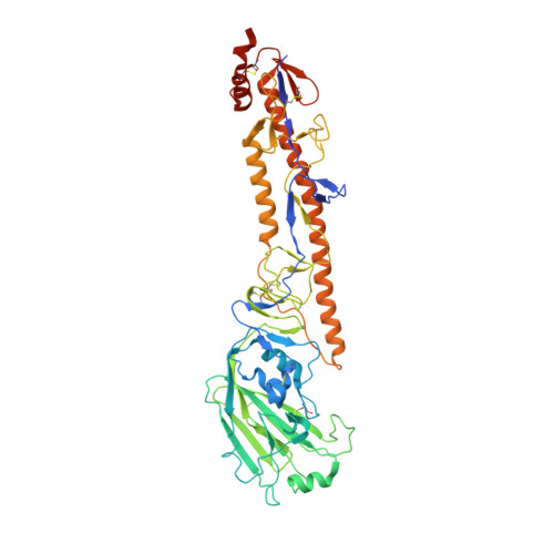
Structural basis for the broad antigenicity of the computationally optimized influenza hemagglutinin X6.

Nagashima, K.A.Dzimianski, J.V.Yang, M.Abendroth, J.Sautto, G.A.Ross, T.M.DuBois, R.M.Edwards, T.E.Mousa, J.J.

(2024) Structure 32: 1079

  • DOI: https://doi.org/10.1016/j.str.2024.05.001
  • Primary Citation Related Structures: 
    8F38, 8GHK, 8SJ9, 8V7O

  • PubMed Abstract: 

    Influenza causes significant morbidity and mortality. As an alternative approach to current seasonal vaccines, the computationally optimized broadly reactive antigen (COBRA) platform has been previously applied to hemagglutinin (HA). This approach integrates wild-type HA sequences into a single immunogen to expand the breadth of accessible antibody epitopes. Adding to previous studies of H1, H3, and H5 COBRA HAs, we define the structural features of another H1 subtype COBRA, X6, that incorporates HA sequences from before and after the 2009 H1N1 influenza pandemic. We determined structures of this antigen alone and in complex with COBRA-specific as well as broadly reactive and functional antibodies, analyzing its antigenicity. We found that X6 possesses features representing both historic and recent H1 HA strains, enabling binding to both head- and stem-reactive antibodies. Overall, these data confirm the integrity of broadly reactive antibody epitopes of X6 and contribute to design efforts for a next-generation vaccine.


  • Organizational Affiliation
    • Center for Vaccines and Immunology, College of Veterinary Medicine, University of Georgia, Athens, GA, USA; Department of Infectious Diseases, College of Veterinary Medicine, University of Georgia, Athens, GA, USA.

Macromolecule Content 

  • Total Structure Weight: 177.73 kDa 
  • Atom Count: 11,845 
  • Modeled Residue Count: 1,455 
  • Deposited Residue Count: 1,539 
  • Unique protein chains: 1

Macromolecules

Find similar proteins by:|  3D Structure
Entity ID: 1
MoleculeChains  Sequence LengthOrganismDetailsImage
Hemagglutinin
A, B, C
513Influenza A virusMutation(s): 0 
Gene Names: HA
UniProt
Find proteins for A0A0G2RXV5 (Influenza A virus)
Explore A0A0G2RXV5 
Go to UniProtKB:  A0A0G2RXV5
Entity Groups
Sequence Clusters30% Identity50% Identity70% Identity90% Identity95% Identity100% Identity
UniProt GroupA0A0G2RXV5
Glycosylation
Glycosylation Sites: 6
Sequence Annotations
Expand
Reference Sequence

Oligosaccharides

Help  
Entity ID: 2
MoleculeChains Length2D Diagram GlycosylationD Interactions
beta-D-mannopyranose-(1-4)-2-acetamido-2-deoxy-beta-D-glucopyranose-(1-4)-2-acetamido-2-deoxy-beta-D-glucopyranose
D, F, G
3N-Glycosylation
Glycosylation Resources
GlyTouCan: G15407YE
GlyCosmos: G15407YE
GlyGen: G15407YE
Entity ID: 3
MoleculeChains Length2D Diagram GlycosylationD Interactions
2-acetamido-2-deoxy-beta-D-glucopyranose-(1-4)-2-acetamido-2-deoxy-beta-D-glucopyranose
E, H
2N-Glycosylation
Glycosylation Resources
GlyTouCan: G42666HT
GlyCosmos: G42666HT
GlyGen: G42666HT

Small Molecules

Ligands 3 Unique
IDChains Name / Formula / InChI Key2D Diagram3D Interactions
NAG

Query on NAG


Download:Ideal Coordinates CCD File
I [auth A]
J [auth A]
K [auth A]
N [auth B]
O [auth B]
I [auth A],
J [auth A],
K [auth A],
N [auth B],
O [auth B],
P [auth B],
Q [auth B],
S [auth C],
T [auth C],
U [auth C]
2-acetamido-2-deoxy-beta-D-glucopyranose
C8 H15 N O6
OVRNDRQMDRJTHS-FMDGEEDCSA-N
GOL

Query on GOL


Download:Ideal Coordinates CCD File
M [auth A]GLYCEROL
C3 H8 O3
PEDCQBHIVMGVHV-UHFFFAOYSA-N
EDO

Query on EDO


Download:Ideal Coordinates CCD File
L [auth A],
R [auth B]
1,2-ETHANEDIOL
C2 H6 O2
LYCAIKOWRPUZTN-UHFFFAOYSA-N

Experimental Data & Validation

Experimental Data

  • Method: X-RAY DIFFRACTION
  • Resolution: 3.25 Å
  • R-Value Free:  0.276 (Depositor), 0.277 (DCC) 
  • R-Value Work:  0.221 (Depositor), 0.221 (DCC) 
  • R-Value Observed: 0.224 (Depositor) 
Space Group: P 1 21 1
Unit Cell:
Length ( Å )Angle ( ˚ )
a = 68.504α = 90
b = 218.725β = 109.68
c = 68.572γ = 90
Software Package:
Software NamePurpose
PHENIXrefinement
XDSdata reduction
Aimlessdata scaling
PHASERphasing
Cootmodel building

Structure Validation

View Full Validation Report


Ligand Structure Quality Assessment 


Entry History 

& Funding Information

Deposition Data


Funding OrganizationLocationGrant Number
National Institutes of Health/National Institute Of Allergy and Infectious Diseases (NIH/NIAID)United States75N93019C00052

Revision History  (Full details and data files)

  • Version 1.0: 2024-05-01
    Type: Initial release
  • Version 1.1: 2024-06-05
    Changes: Database references
  • Version 1.2: 2024-08-21
    Changes: Database references
  • Version 1.3: 2024-08-28
    Changes: Database references
  • Version 1.4: 2024-10-30
    Changes: Structure summary