8SIP | pdb_00008sip

Structure of a mouse IgG antibody fragment that binds Inosine, an RNA modification


Experimental Data Snapshot

  • Method: X-RAY DIFFRACTION
  • Resolution: 1.94 Å
  • R-Value Free: 
    0.245 (Depositor), 0.245 (DCC) 
  • R-Value Work: 
    0.204 (Depositor), 0.204 (DCC) 
  • R-Value Observed: 
    0.206 (Depositor) 

Starting Model: experimental
View more details

wwPDB Validation 3D Report Full Report

Validation slider image for 8SIP

Ligand Structure Quality Assessment 


This is version 1.2 of the entry. See complete history

Literature

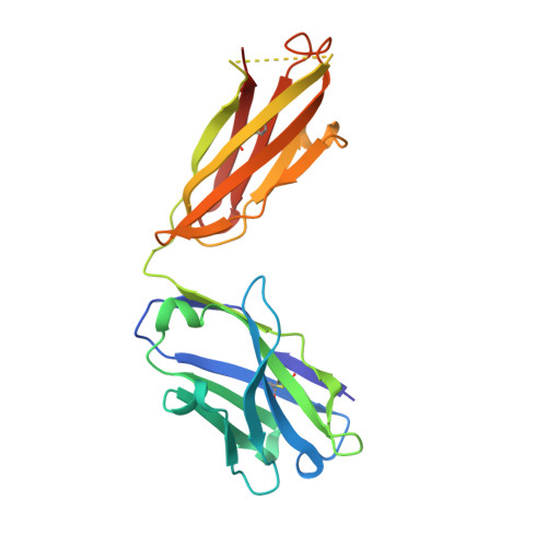
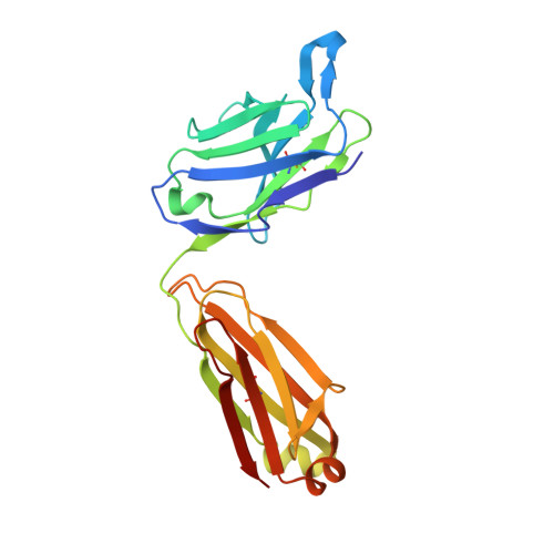
In silico lambda-dynamics predicts protein binding specificities to modified RNAs.

Angelo, M.Zhang, W.Vilseck, J.Z.Aoki, S.T.

(2025) Nucleic Acids Res 53

  • DOI: https://doi.org/10.1093/nar/gkaf166
  • Primary Citation Related Structures: 
    8SIP, 8TCA, 8VEV

  • PubMed Abstract: 

    RNA modifications shape gene expression through a variety of chemical changes to canonical RNA bases. Although numbering in the hundreds, only a few RNA modifications are well characterized, in part due to the absence of methods to identify modification sites. Antibodies remain a common tool to identify modified RNA and infer modification sites through straightforward applications. However, specificity issues can result in off-target binding and confound conclusions. This work utilizes in silico λ-dynamics to efficiently estimate binding free energy differences of modification-targeting antibodies between a variety of naturally occurring RNA modifications. Crystal structures of inosine and N6-methyladenosine (m6A) targeting antibodies bound to their modified ribonucleosides were determined and served as structural starting points. λ-Dynamics was utilized to predict RNA modifications that permit or inhibit binding to these antibodies. In vitro RNA-antibody binding assays supported the accuracy of these in silico results. High agreement between experimental and computed binding propensities demonstrated that λ-dynamics can serve as a predictive screen for antibody specificity against libraries of RNA modifications. More importantly, this strategy is an innovative way to elucidate how hundreds of known RNA modifications interact with biological molecules without the limitations imposed by in vitro or in vivo methodologies.


  • Organizational Affiliation
    • Department of Biochemistry and Molecular Biology, Indiana University School of Medicine, 635 Barnhill Drive, Indianapolis, IN 46202, United States.

Macromolecule Content 

  • Total Structure Weight: 48.51 kDa 
  • Atom Count: 3,531 
  • Modeled Residue Count: 426 
  • Deposited Residue Count: 441 
  • Unique protein chains: 2

Macromolecules

Find similar proteins by:|  3D Structure
Entity ID: 1
MoleculeChains  Sequence LengthOrganismDetailsImage
Inosine binding IgG Fab, heavy chainA [auth H]222Mus musculusMutation(s): 0 
Find similar proteins by:|  3D Structure
Entity ID: 2
MoleculeChains  Sequence LengthOrganismDetailsImage
Inosine binding IgG Fab, kappa chainB [auth L]219Mus musculusMutation(s): 0 

Small Molecules

Ligands 1 Unique
IDChains Name / Formula / InChI Key2D Diagram3D Interactions
NOS
(Subject of Investigation/LOI)

Query on NOS


Download:Ideal Coordinates CCD File
C [auth H]INOSINE
C10 H12 N4 O5
UGQMRVRMYYASKQ-KQYNXXCUSA-N

Experimental Data & Validation

Experimental Data

  • Method: X-RAY DIFFRACTION
  • Resolution: 1.94 Å
  • R-Value Free:  0.245 (Depositor), 0.245 (DCC) 
  • R-Value Work:  0.204 (Depositor), 0.204 (DCC) 
  • R-Value Observed: 0.206 (Depositor) 
Space Group: P 1
Unit Cell:
Length ( Å )Angle ( ˚ )
a = 39.817α = 83.84
b = 49.09β = 88.82
c = 57.385γ = 89.68
Software Package:
Software NamePurpose
PHENIXrefinement
autoPROCdata processing
Aimlessdata scaling
PHASERphasing
Cootmodel building
autoPROCdata reduction

Structure Validation

View Full Validation Report


Ligand Structure Quality Assessment 


Entry History 

& Funding Information

Deposition Data

  • Released Date: 2024-02-07 
  • Deposition Author(s): Aoki, S.T.

Funding OrganizationLocationGrant Number
Not funded--

Revision History  (Full details and data files)

  • Version 1.0: 2024-02-07
    Type: Initial release
  • Version 1.1: 2024-11-06
    Changes: Structure summary
  • Version 1.2: 2025-08-20
    Changes: Database references