8SIC | pdb_00008sic

Crystal structure of Epstein-Barr virus glycoprotein 350 (gp350) in complex with Cy137C02, a monoclonal antibody isolated from macaques immunized with a gp350 nanoparticle vaccine


Experimental Data Snapshot

  • Method: X-RAY DIFFRACTION
  • Resolution: 2.76 Å
  • R-Value Free: 
    0.295 (Depositor), 0.296 (DCC) 
  • R-Value Work: 
    0.245 (Depositor), 0.252 (DCC) 
  • R-Value Observed: 
    0.248 (Depositor) 

Starting Model: experimental
View more details

wwPDB Validation 3D Report Full Report

Validation slider image for 8SIC

Ligand Structure Quality Assessment 


This is version 1.4 of the entry. See complete history

Literature

Structural basis for complement receptor engagement and virus neutralization through Epstein-Barr virus gp350.

Joyce, M.G.Bu, W.Chen, W.H.Gillespie, R.A.Andrews, S.F.Wheatley, A.K.Tsybovsky, Y.Jensen, J.L.Stephens, T.Prabhakaran, M.Fisher, B.E.Narpala, S.R.Bagchi, M.McDermott, A.B.Nabel, G.J.Kwong, P.D.Mascola, J.R.Cohen, J.I.Kanekiyo, M.

(2025) Immunity 58: 295

  • DOI: https://doi.org/10.1016/j.immuni.2025.01.010
  • Primary Citation Related Structures: 
    8SEF, 8SGA, 8SGG, 8SGN, 8SIC, 8SM0, 8SM1

  • PubMed Abstract: 

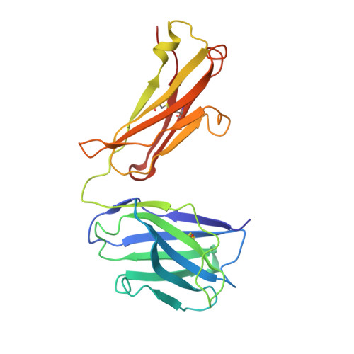
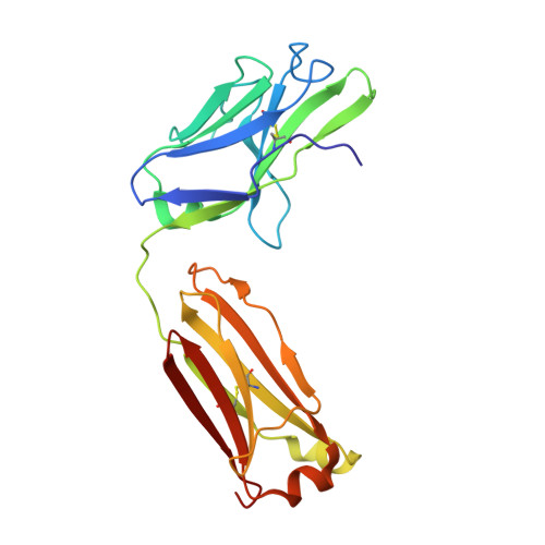
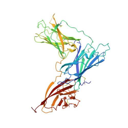
    Epstein-Barr virus (EBV) causes infectious mononucleosis and is associated with malignancies in humans. Viral infection of B cells is initiated by the viral glycoprotein 350 (gp350) binding to complement receptor 2 (CR2). Despite decades of effort, no vaccines or curative agents have been developed, partly due to lack of atomic-level understanding of the virus-host interface. Here, we determined the 1.7 Å structure of gp350 in complex with CR2. CR2 binding of gp350 utilized the same set of Arg residues required for recognition of its natural ligand, complement C3d. We further determined the structures of gp350 in complex with three potently neutralizing antibodies (nAbs) obtained from vaccinated macaques and EBV-infected individuals. Like the CR2 interaction, these nAbs targeted the acidic pocket within the CR2-binding site on gp350 using Arg residues. Our results illustrate two axes of molecular mimicry-gp350 versus C3d and CR2 versus EBV nAbs-offering insights for EBV vaccines and therapeutics development.


  • Organizational Affiliation
    • Vaccine Research Center, National Institute of Allergy and Infectious Diseases, National Institutes of Health, Bethesda, MD 20892, USA; Henry M. Jackson Foundation for the Advancement of Military Medicine, Inc, Bethesda, MD 20817, USA; Emerging Infectious Diseases Branch, Walter Reed Army Institute of Research, Silver Spring, MD 20910, USA. Electronic address: gjoyce@eidresearch.org.

Macromolecule Content 

  • Total Structure Weight: 190.91 kDa 
  • Atom Count: 13,230 
  • Modeled Residue Count: 1,696 
  • Deposited Residue Count: 1,736 
  • Unique protein chains: 3

Macromolecules

Find similar proteins by:|  3D Structure
Entity ID: 1
MoleculeChains  Sequence LengthOrganismDetailsImage
Cy137C02 Fab heavy chainA,
E [auth H]
222Macaca fascicularisMutation(s): 0 
Find similar proteins by:|  3D Structure
Entity ID: 2
MoleculeChains  Sequence LengthOrganismDetailsImage
Cy137C02 Fab light chainB,
F [auth L]
215Macaca fascicularisMutation(s): 0 
Find similar proteins by:|  3D Structure
Entity ID: 3
MoleculeChains  Sequence LengthOrganismDetailsImage
Envelope glycoprotein gp350C [auth E],
D [auth G]
431Human herpesvirus 4 strain B95-8Mutation(s): 0 
Gene Names: BLLF1
UniProt
Find proteins for P03200 (Epstein-Barr virus (strain B95-8))
Explore P03200 
Go to UniProtKB:  P03200
Entity Groups
Sequence Clusters30% Identity50% Identity70% Identity90% Identity95% Identity100% Identity
UniProt GroupP03200
Glycosylation
Glycosylation Sites: 9
Sequence Annotations
Expand
Reference Sequence

Small Molecules

Ligands 2 Unique
IDChains Name / Formula / InChI Key2D Diagram3D Interactions
NAG

Query on NAG


Download:Ideal Coordinates CCD File
G [auth E]
H [auth E]
I [auth E]
J [auth E]
K [auth E]
G [auth E],
H [auth E],
I [auth E],
J [auth E],
K [auth E],
L [auth E],
N [auth E],
O [auth E],
P [auth G],
Q [auth G],
R [auth G],
S [auth G],
T [auth G],
U [auth G],
V [auth G],
W [auth G],
X [auth G]
2-acetamido-2-deoxy-beta-D-glucopyranose
C8 H15 N O6
OVRNDRQMDRJTHS-FMDGEEDCSA-N
ZN

Query on ZN


Download:Ideal Coordinates CCD File
M [auth E],
Y [auth G]
ZINC ION
Zn
PTFCDOFLOPIGGS-UHFFFAOYSA-N

Experimental Data & Validation

Experimental Data

  • Method: X-RAY DIFFRACTION
  • Resolution: 2.76 Å
  • R-Value Free:  0.295 (Depositor), 0.296 (DCC) 
  • R-Value Work:  0.245 (Depositor), 0.252 (DCC) 
  • R-Value Observed: 0.248 (Depositor) 
Space Group: P 21 2 21
Unit Cell:
Length ( Å )Angle ( ˚ )
a = 69.047α = 90
b = 71.377β = 90
c = 395.083γ = 90
Software Package:
Software NamePurpose
PHENIXrefinement
HKL-2000data reduction
HKL-2000data scaling
PHASERphasing

Structure Validation

View Full Validation Report


Ligand Structure Quality Assessment 


Entry History 

& Funding Information

Deposition Data


Funding OrganizationLocationGrant Number
Department of Defense (DOD, United States)United StatesW81XWH-07-2-0067

Revision History  (Full details and data files)

  • Version 1.0: 2024-04-17
    Type: Initial release
  • Version 1.1: 2024-11-13
    Changes: Structure summary
  • Version 1.2: 2025-02-12
    Changes: Database references
  • Version 1.3: 2025-02-19
    Changes: Database references
  • Version 1.4: 2025-02-26
    Changes: Database references