8S6O | pdb_00008s6o

Structure of MLLE3 domain of Rrm4 in complex with PAM2L2 of Upa1


Experimental Data Snapshot

  • Method: X-RAY DIFFRACTION
  • Resolution: 2.40 Å
  • R-Value Free: 
    0.276 (Depositor), 0.269 (DCC) 
  • R-Value Work: 
    0.240 (Depositor), 0.233 (DCC) 

Starting Model: experimental
View more details

wwPDB Validation 3D Report Full Report

Validation slider image for 8S6O

This is version 1.2 of the entry. See complete history

Literature

Deciphering the RNA-binding protein network during endosomal mRNA transport.

Devan, S.K.Shanmugasundaram, S.Muntjes, K.Postma, J.Smits, S.H.J.Altegoer, F.Feldbrugge, M.

(2024) Proc Natl Acad Sci U S A 121: e2404091121-e2404091121

  • DOI: https://doi.org/10.1073/pnas.2404091121
  • Primary Citation Related Structures: 
    8S6N, 8S6O, 8S6U

  • PubMed Abstract: 

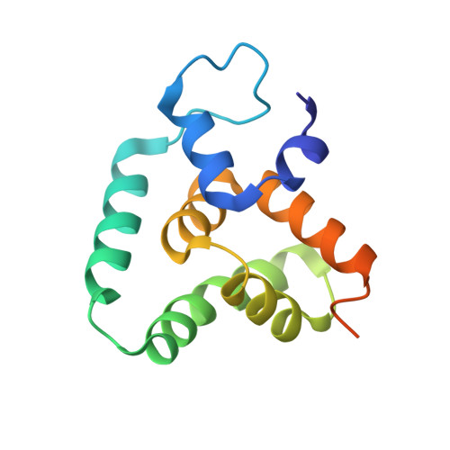
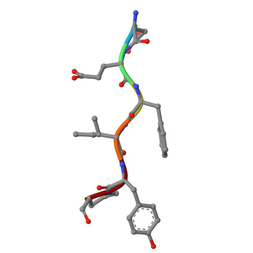
    Microtubule-dependent endosomal transport is crucial for polar growth, ensuring the precise distribution of cellular cargos such as proteins and mRNAs. However, the molecular mechanism linking mRNAs to the endosomal surface remains poorly understood. Here, we present a structural analysis of the key RNA-binding protein Rrm4 from Ustilago maydis . Our findings reveal a different type of MademoiseLLE domain (MLLE) featuring a seven-helical bundle that provides a distinct binding interface. A comparative analysis with the canonical MademoiseLLE domain of the poly(A)-binding protein Pab1 disclosed unique characteristics of both domains. Deciphering the MLLE binding code enabled prediction and verification of previously unknown Rrm4 interactors containing short linear motifs. Importantly, we demonstrated that the human MLLE domains, such as those of PABPC1 and UBR5, employed a similar principle to distinguish among interaction partners. Thus, our study provides detailed mechanistic insights into how structural variations in the widely distributed MLLE domain facilitate mRNA attachment during endosomal transport.


  • Organizational Affiliation
    • Department of Biology, Institute of Microbiology, Cluster of Excellence on Plant Sciences, Heinrich Heine University Düsseldorf, Düsseldorf 40204, Germany.

Macromolecule Content 

  • Total Structure Weight: 121.61 kDa 
  • Atom Count: 7,457 
  • Modeled Residue Count: 968 
  • Deposited Residue Count: 1,112 
  • Unique protein chains: 2

Macromolecules

Find similar proteins by:|  3D Structure
Entity ID: 1
MoleculeChains  Sequence LengthOrganismDetailsImage
RNA-binding protein RRM4133Mycosarcoma maydisMutation(s): 0 
Gene Names: RRM4UMAG_10836
UniProt
Find proteins for A0A0D1DWZ5 (Mycosarcoma maydis)
Explore A0A0D1DWZ5 
Go to UniProtKB:  A0A0D1DWZ5
Entity Groups
Sequence Clusters30% Identity50% Identity70% Identity90% Identity95% Identity100% Identity
UniProt GroupA0A0D1DWZ5
Sequence Annotations
Expand
Reference Sequence
Find similar proteins by:  Sequence   |   3D Structure  
Entity ID: 2
MoleculeChains  Sequence LengthOrganismDetailsImage
PAMPL26Mycosarcoma maydisMutation(s): 0 

Experimental Data & Validation

Experimental Data

  • Method: X-RAY DIFFRACTION
  • Resolution: 2.40 Å
  • R-Value Free:  0.276 (Depositor), 0.269 (DCC) 
  • R-Value Work:  0.240 (Depositor), 0.233 (DCC) 
Space Group: C 1 2 1
Unit Cell:
Length ( Å )Angle ( ˚ )
a = 82.881α = 90
b = 74.633β = 90.08
c = 168.511γ = 90
Software Package:
Software NamePurpose
PHENIXrefinement
XDSdata reduction
XSCALEdata scaling
PHASERphasing

Structure Validation

View Full Validation Report


Entry History 

& Funding Information

Deposition Data


Funding OrganizationLocationGrant Number
German Research Foundation (DFG)Germany417919780

Revision History  (Full details and data files)

  • Version 1.0: 2024-10-30
    Type: Initial release
  • Version 1.1: 2024-11-13
    Changes: Database references
  • Version 1.2: 2024-11-20
    Changes: Structure summary