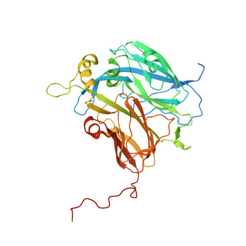
8S5X

High pH (8.0) as-isolated MSOX movie series dataset 10 of the copper nitrite reductase (NirK) from Bradyrhizobium japonicum USDA110 [3.5 MGy]


Experimental Data Snapshot

  • Method: X-RAY DIFFRACTION
  • Resolution: 1.12 Å
  • R-Value Free: 0.133 
  • R-Value Work: 0.113 

Starting Model: experimental
View more details

wwPDB Validation   3D Report Full Report


This is version 1.1 of the entry. See complete history


Literature

Spectroscopically Validated pH-dependent MSOX Movies Provide Detailed Mechanism of Copper Nitrite Reductases.

Rose, S.L.Ferroni, F.M.Horrell, S.Brondino, C.D.Eady, R.R.Jaho, S.Hough, M.A.Owen, R.L.Antonyuk, S.V.Hasnain, S.S.

(2024) J Mol Biol 436: 168706-168706

  • DOI: https://doi.org/10.1016/j.jmb.2024.168706
  • Primary Citation of Related Structures:  
    8R8S, 8RFL, 8RFO, 8RFP, 8RFQ, 8RFR, 8RFS, 8RFT, 8RFU, 8RFV, 8RFW, 8RFX, 8RFY, 8RG8, 8RG9, 8RGB, 8RGC, 8RGD, 8RU9, 8RUR, 8RYJ, 8RYR, 8RYU, 8RYV, 8S0W, 8S2Q, 8S5X, 8S5Y, 8S63, 8S64, 8S68, 8S69, 9EVM

  • PubMed Abstract: 

    Copper nitrite reductases (CuNiRs) exhibit a strong pH dependence of their catalytic activity. Structural movies can be obtained by serially recording multiple structures (frames) from the same spot of a crystal using the MSOX serial crystallography approach. This method has been combined with on-line single crystal optical spectroscopy to capture the pH-dependent structural changes that accompany during turnover of CuNiRs from two Rhizobia species. The structural movies, initiated by the redox activation of a type-1 copper site (T1Cu) via X-ray generated photoelectrons, have been obtained for the substrate-free and substrate-bound states at low (high enzymatic activity) and high (low enzymatic activity) pH. At low pH, formation of the product nitric oxide (NO) is complete at the catalytic type-2 copper site (T2Cu) after a dose of 3 MGy (frame 5) with full bleaching of the T1Cu ligand-to-metal charge transfer (LMCT) 455 nm band (S(σ) Cys → T1Cu 2+ ) which in itself indicates the electronic route of proton-coupled electron transfer (PCET) from T1Cu to T2Cu. In contrast at high pH, the changes in optical spectra are relatively small and the formation of NO is only observed in later frames (frame 15 in Br 2D NiR, 10 MGy), consistent with the loss of PCET required for catalysis. This is accompanied by decarboxylation of the catalytic Asp CAT residue, with CO 2 trapped in the catalytic pocket.


  • Organizational Affiliation

    Molecular Biophysics Group, Life Sciences Building, Institute of Systems, Molecular and Integrative Biology, Faculty of Health and Life Sciences, University of Liverpool, Liverpool, L69 7ZB, United Kingdom.


Macromolecules
Find similar proteins by:  (by identity cutoff)  |  3D Structure
Entity ID: 1
MoleculeChains Sequence LengthOrganismDetailsImage
Copper-containing nitrite reductase367Bradyrhizobium sp.Mutation(s): 0 
Gene Names: nirK
EC: 1.7.2.1
UniProt
Find proteins for Q89EJ6 (Bradyrhizobium diazoefficiens (strain JCM 10833 / BCRC 13528 / IAM 13628 / NBRC 14792 / USDA 110))
Explore Q89EJ6 
Go to UniProtKB:  Q89EJ6
Entity Groups  
Sequence Clusters30% Identity50% Identity70% Identity90% Identity95% Identity100% Identity
UniProt GroupQ89EJ6
Sequence Annotations
Expand
  • Reference Sequence
Small Molecules
Ligands 4 Unique
IDChains Name / Formula / InChI Key2D Diagram3D Interactions
FRU
Query on FRU

Download Ideal Coordinates CCD File 
E [auth A]beta-D-fructofuranose
C6 H12 O6
RFSUNEUAIZKAJO-ARQDHWQXSA-N
GLC
Query on GLC

Download Ideal Coordinates CCD File 
D [auth A]alpha-D-glucopyranose
C6 H12 O6
WQZGKKKJIJFFOK-DVKNGEFBSA-N
SO4
Query on SO4

Download Ideal Coordinates CCD File 
F [auth A]
G [auth A]
H [auth A]
I [auth A]
J [auth A]
F [auth A],
G [auth A],
H [auth A],
I [auth A],
J [auth A],
K [auth A]
SULFATE ION
O4 S
QAOWNCQODCNURD-UHFFFAOYSA-L
CU (Subject of Investigation/LOI)
Query on CU

Download Ideal Coordinates CCD File 
B [auth A],
C [auth A]
COPPER (II) ION
Cu
JPVYNHNXODAKFH-UHFFFAOYSA-N
Experimental Data & Validation

Experimental Data

  • Method: X-RAY DIFFRACTION
  • Resolution: 1.12 Å
  • R-Value Free: 0.133 
  • R-Value Work: 0.113 
  • Space Group: P 63
Unit Cell:
Length ( Å )Angle ( ˚ )
a = 103.97α = 90
b = 103.97β = 90
c = 64.46γ = 120
Software Package:
Software NamePurpose
REFMACrefinement
REFMACrefinement
xia2data reduction
Aimlessdata scaling
REFMACphasing

Structure Validation

View Full Validation Report


Entry History & Funding Information

Deposition Data


Funding OrganizationLocationGrant Number
Agencia Nacional de Promocion Cientifica y Tecnologica (FONCYT)ArgentinaPICT 2020-00522
Biotechnology and Biological Sciences Research Council (BBSRC)United KingdomBB/N013972/1

Revision History  (Full details and data files)

  • Version 1.0: 2024-07-24
    Type: Initial release
  • Version 1.1: 2024-08-07
    Changes: Database references, Structure summary