8RYI

Metformin hydrolase from Aminobacter niigataensis MD1 with urea in the active site


Experimental Data Snapshot

  • Method: X-RAY DIFFRACTION
  • Resolution: 2.06 Å
  • R-Value Free: 0.199 
  • R-Value Work: 0.169 
  • R-Value Observed: 0.170 

Starting Model: in silico
View more details

wwPDB Validation   3D Report Full Report


Ligand Structure Quality Assessment 


This is version 1.1 of the entry. See complete history


Literature

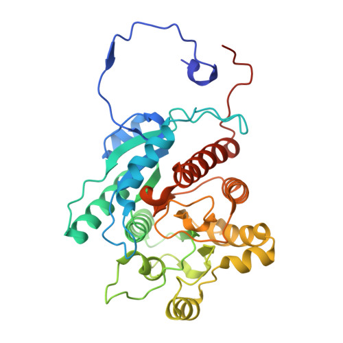
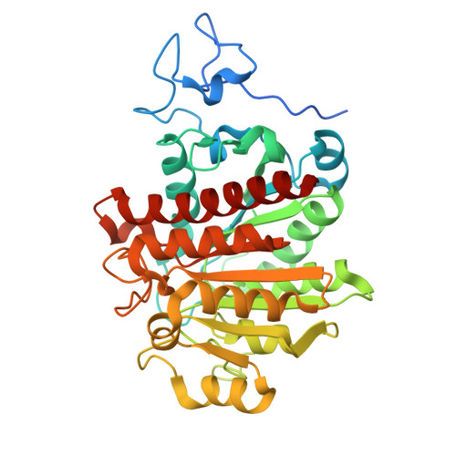
Metformin hydrolase is a recently evolved nickel-dependent heteromeric ureohydrolase.

Sinn, M.Riede, L.Fleming, J.R.Funck, D.Lutz, H.Bachmann, A.Mayans, O.Hartig, J.S.

(2024) Nat Commun 15: 8045-8045

  • DOI: https://doi.org/10.1038/s41467-024-51752-5
  • Primary Citation of Related Structures:  
    8RYI

  • PubMed Abstract: 

    The anti-diabetic drug metformin is one of the most widely prescribed medicines in the world. Together with its degradation product guanylurea, it is a major pharmaceutical pollutant in wastewater treatment plants and surface waters. An operon comprising two genes of the ureohydrolase family in Pseudomonas and Aminobacter species has recently been implicated in metformin degradation. However, the corresponding proteins have not been characterized. Here we show that these genes encode a Ni 2+ -dependent enzyme that efficiently and specifically hydrolyzes metformin to guanylurea and dimethylamine. The active enzyme is a heteromeric complex of α- and β- subunits in which only the α-subunits contain the conserved His and Asp residues for the coordination of two Ni 2+ ions in the active site. A crystal structure of metformin hydrolase reveals an α 2 β 4 stoichiometry of the hexameric complex, which is unprecedented in the ureohydrolase family. By studying a closely related but more widely distributed enzyme, we find that the putative predecessor specifically hydrolyzes dimethylguanidine instead of metformin. Our findings establish the molecular basis for metformin hydrolysis to guanylurea as the primary pathway for metformin biodegradation and provide insight into the recent evolution of ureohydrolase family proteins in response to an anthropogenic compound.


  • Organizational Affiliation

    Department of Chemistry, University of Konstanz, Konstanz, Germany. malte.sinn@uni-konstanz.de.


Macromolecules
Find similar proteins by:  (by identity cutoff)  |  3D Structure
Entity ID: 1
MoleculeChains Sequence LengthOrganismDetailsImage
Arginase family proteinA [auth C],
C [auth A],
E [auth F],
F [auth D]
348Aminobacter niigataensisMutation(s): 0 
Gene Names: N7E70_023835
Entity Groups  
Sequence Clusters30% Identity50% Identity70% Identity90% Identity95% Identity100% Identity
Sequence Annotations
Expand
  • Reference Sequence
Find similar proteins by:  (by identity cutoff)  |  3D Structure
Entity ID: 2
MoleculeChains Sequence LengthOrganismDetailsImage
Agmatinase family proteinB,
D [auth E]
376Aminobacter niigataensisMutation(s): 0 
Gene Names: N7E70_023840
Entity Groups  
Sequence Clusters30% Identity50% Identity70% Identity90% Identity95% Identity100% Identity
Sequence Annotations
Expand
  • Reference Sequence
Small Molecules
Ligands 4 Unique
IDChains Name / Formula / InChI Key2D Diagram3D Interactions
C5J
Query on C5J

Download Ideal Coordinates CCD File 
G [auth B],
K [auth E]
dicarbonimidic diamide
C2 H5 N3 O2
OHJMTUPIZMNBFR-UHFFFAOYSA-N
URE (Subject of Investigation/LOI)
Query on URE

Download Ideal Coordinates CCD File 
H [auth B],
L [auth E]
UREA
C H4 N2 O
XSQUKJJJFZCRTK-UHFFFAOYSA-N
NI (Subject of Investigation/LOI)
Query on NI

Download Ideal Coordinates CCD File 
I [auth B],
J [auth B],
M [auth E],
N [auth E]
NICKEL (II) ION
Ni
VEQPNABPJHWNSG-UHFFFAOYSA-N
CA
Query on CA

Download Ideal Coordinates CCD File 
O [auth E]CALCIUM ION
Ca
BHPQYMZQTOCNFJ-UHFFFAOYSA-N
Experimental Data & Validation

Experimental Data

  • Method: X-RAY DIFFRACTION
  • Resolution: 2.06 Å
  • R-Value Free: 0.199 
  • R-Value Work: 0.169 
  • R-Value Observed: 0.170 
  • Space Group: C 1 2 1
Unit Cell:
Length ( Å )Angle ( ˚ )
a = 163.99α = 90
b = 86.52β = 113.99
c = 168.41γ = 90
Software Package:
Software NamePurpose
PHENIXrefinement
XDSdata reduction
XSCALEdata scaling
PHASERphasing

Structure Validation

View Full Validation Report


Ligand Structure Quality Assessment 


Entry History & Funding Information

Deposition Data


Funding OrganizationLocationGrant Number
European Research Council (ERC)European Union681777

Revision History  (Full details and data files)

  • Version 1.0: 2024-08-28
    Type: Initial release
  • Version 1.1: 2024-10-02
    Changes: Database references