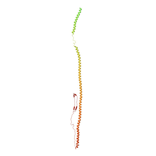
Vimentin filaments integrate low-complexity domains in a complex helical structure.
Eibauer, M., Weber, M.S., Kronenberg-Tenga, R., Beales, C.T., Boujemaa-Paterski, R., Turgay, Y., Sivagurunathan, S., Kraxner, J., Koster, S., Goldman, R.D., Medalia, O.(2024) Nat Struct Mol Biol 31: 939-949
- PubMed: 38632361 Search on PubMedSearch on PubMed Central
- DOI: https://doi.org/10.1038/s41594-024-01261-2
- Primary Citation Related Structures:
8RVE - PubMed Abstract:
Intermediate filaments (IFs) are integral components of the cytoskeleton. They provide cells with tissue-specific mechanical properties and are involved in numerous cellular processes. Due to their intricate architecture, a 3D structure of IFs has remained elusive. Here we use cryo-focused ion-beam milling, cryo-electron microscopy and tomography to obtain a 3D structure of vimentin IFs (VIFs). VIFs assemble into a modular, intertwined and flexible helical structure of 40 α-helices in cross-section, organized into five protofibrils. Surprisingly, the intrinsically disordered head domains form a fiber in the lumen of VIFs, while the intrinsically disordered tails form lateral connections between the protofibrils. Our findings demonstrate how protein domains of low sequence complexity can complement well-folded protein domains to construct a biopolymer with striking mechanical strength and stretchability.
- Department of Biochemistry, University of Zurich, Zurich, Switzerland. m.eibauer@bioc.uzh.ch.
Organizational Affiliation: