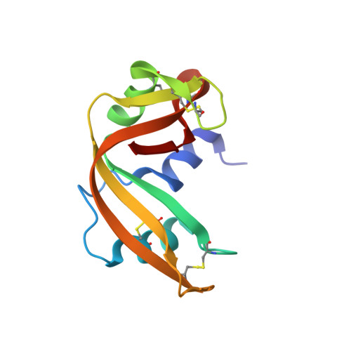
8RSA

CRYSTAL STRUCTURE OF TWO COVALENT NUCLEOSIDE DERIVATIVES OF RIBONUCLEASE A


Experimental Data Snapshot

  • Method: X-RAY DIFFRACTION
  • Resolution: 1.80 Å
  • R-Value Observed: 0.162 

wwPDB Validation   3D Report Full Report


This is version 1.4 of the entry. See complete history


Literature

Crystal structure of two covalent nucleoside derivatives of ribonuclease A.

Nachman, J.Miller, M.Gilliland, G.L.Carty, R.Pincus, M.Wlodawer, A.

(1990) Biochemistry 29: 928-937

  • DOI: https://doi.org/10.1021/bi00456a012
  • Primary Citation of Related Structures:  
    8RSA, 9RSA

  • PubMed Abstract: 

    Crystal structures of two forms of ribonuclease A with deoxynucleosides covalently bound to respectively His 12 and His 119 have been solved. One form, T-H12-RNase, has a deoxythymidine bound to N epsilon 2 of His 12, while the other one, U-H119-RNase, has a deoxyuridine bound to N delta 1 of His 119. The two crystal forms are nearly isomorphous, with two molecules in the asymmetric unit. However, the modified ribonucleases differ both in their enzymatic activities and in the conformation of the catalytic site and of the deoxynucleoside-histidine moiety. T-H12-RNase is characterized by complete loss of enzymatic activity; in this form the deoxynucleoside completely blocks the catalytic site and forms intramolecular contacts with residues associated with both the B1 and B2 sites. U-H119-RNase retains 1% of the activity of the unmodified enzyme, and in this form His 119 adopts a different orientation, corresponding to the alternate conformation reported for this residue; the deoxynucleoside-histidine moiety points out of the active site and does not form any contacts with the rest of the protein, thus allowing partial access to the catalytic site. On the basis of these structures, we propose possible mechanisms for the reactions of bromoacetamido nucleosides with ribonuclease A.


  • Organizational Affiliation

    BRI Basic Research Program, NCI-Frederick Cancer Research Facility, Maryland 21701.


Macromolecules
Find similar proteins by:  (by identity cutoff)  |  3D Structure
Entity ID: 1
MoleculeChains Sequence LengthOrganismDetailsImage
RIBONUCLEASE A
A, B
124Bos taurusMutation(s): 0 
EC: 3.1.27.5 (PDB Primary Data), 4.6.1.18 (UniProt)
UniProt
Find proteins for P61823 (Bos taurus)
Explore P61823 
Go to UniProtKB:  P61823
Entity Groups  
Sequence Clusters30% Identity50% Identity70% Identity90% Identity95% Identity100% Identity
UniProt GroupP61823
Sequence Annotations
Expand
  • Reference Sequence
Small Molecules
Ligands 1 Unique
IDChains Name / Formula / InChI Key2D Diagram3D Interactions
ADT
Query on ADT

Download Ideal Coordinates CCD File 
C [auth A],
D [auth B]
3'-DEOXY-3'-ACETAMIDO-THYMIDINE
C12 H17 N3 O5
OCWNRLPPKHGFCE-IVZWLZJFSA-N
Experimental Data & Validation

Experimental Data

  • Method: X-RAY DIFFRACTION
  • Resolution: 1.80 Å
  • R-Value Observed: 0.162 
  • Space Group: P 21 21 21
Unit Cell:
Length ( Å )Angle ( ˚ )
a = 53.14α = 90
b = 64.61β = 90
c = 73.64γ = 90
Software Package:
Software NamePurpose
PROFFTrefinement

Structure Validation

View Full Validation Report


Entry History 

Deposition Data

Revision History  (Full details and data files)

  • Version 1.0: 1991-04-15
    Type: Initial release
  • Version 1.1: 2008-03-03
    Changes: Version format compliance
  • Version 1.2: 2011-07-13
    Changes: Version format compliance
  • Version 1.3: 2024-06-05
    Changes: Data collection, Database references, Derived calculations, Other
  • Version 1.4: 2024-11-06
    Changes: Structure summary