Low-dose cryo-electron ptychography of proteins at sub-nanometer resolution.
Kucukoglu, B., Mohammed, I., Guerrero-Ferreira, R.C., Ribet, S.M., Varnavides, G., Leidl, M.L., Lau, K., Nazarov, S., Myasnikov, A., Kube, M., Radecke, J., Sachse, C., Muller-Caspary, K., Ophus, C., Stahlberg, H.(2024) Nat Commun 15: 8062-8062
- PubMed: 39277607 Search on PubMedSearch on PubMed Central
- DOI: https://doi.org/10.1038/s41467-024-52403-5
- Primary Citation Related Structures:
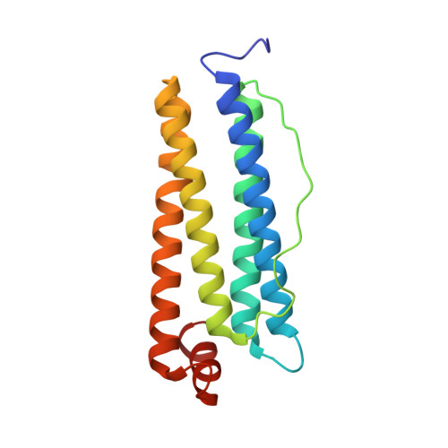
8RQB - PubMed Abstract:
Cryo-transmission electron microscopy (cryo-EM) of frozen hydrated specimens is an efficient method for the structural analysis of purified biological molecules. However, cryo-EM and cryo-electron tomography are limited by the low signal-to-noise ratio (SNR) of recorded images, making detection of smaller particles challenging. For dose-resilient samples often studied in the physical sciences, electron ptychography - a coherent diffractive imaging technique using 4D scanning transmission electron microscopy (4D-STEM) - has recently demonstrated excellent SNR and resolution down to tens of picometers for thin specimens imaged at room temperature. Here we apply 4D-STEM and ptychographic data analysis to frozen hydrated proteins, reaching sub-nanometer resolution 3D reconstructions. We employ low-dose cryo-EM with an aberration-corrected, convergent electron beam to collect 4D-STEM data for our reconstructions. The high frame rate of the electron detector allows us to record large datasets of electron diffraction patterns with substantial overlaps between the interaction volumes of adjacent scan positions, from which the scattering potentials of the samples are iteratively reconstructed. The reconstructed micrographs show strong SNR enabling the reconstruction of the structure of apoferritin protein at up to 5.8 Å resolution. We also show structural analysis of the Phi92 capsid and sheath, tobacco mosaic virus, and bacteriorhodopsin at slightly lower resolutions.
- Laboratory of Biological Electron Microscopy, Institute of Physics, School of Basic Sciences, EPFL, and Department of Fundamental Microbiology, Faculty of Biology and Medicine, UNIL, Rte. de la Sorge, 1015, Lausanne, Switzerland.
Organizational Affiliation: