8RPT | pdb_00008rpt

Crystal structure of human DNPH1 mutant- D80A


Experimental Data Snapshot

  • Method: X-RAY DIFFRACTION
  • Resolution: 1.95 Å
  • R-Value Free: 
    0.220 (Depositor), 0.221 (DCC) 
  • R-Value Work: 
    0.178 (Depositor), 0.184 (DCC) 
  • R-Value Observed: 
    0.180 (Depositor) 

Starting Model: experimental
View more details

wwPDB Validation 3D Report Full Report

Validation slider image for 8RPT

Ligand Structure Quality Assessment 


This is version 1.0 of the entry. See complete history

Literature

Human 2'-Deoxynucleoside 5'-Phosphate N-Hydrolase 1: The Catalytic Roles of Tyr24 and Asp80.

Carberry, A.E.Devi, S.Harrison, D.J.da Silva, R.G.

(2024) Chembiochem 25: e202400047-e202400047

  • DOI: https://doi.org/10.1002/cbic.202400047
  • Primary Citation Related Structures: 
    8RPS, 8RPT, 8RQD

  • PubMed Abstract: 

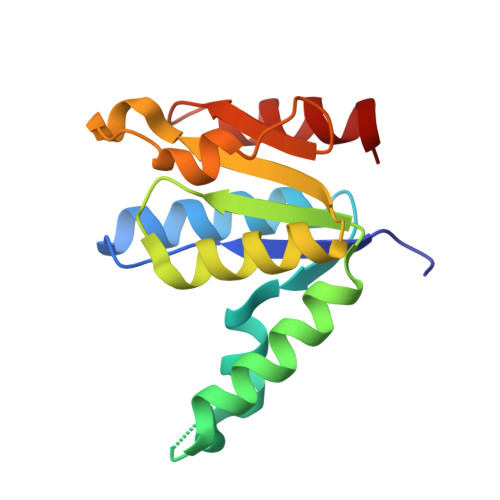
    The human enzyme 2'-deoxynucleoside 5'-phosphate N-hydrolase 1 (HsDNPH1) catalyses the hydrolysis of 5-hydroxymethyl-2'-deoxyuridine 5'-phosphate to generate 5-hydroxymethyluracil and 2-deoxyribose-5-phosphate via a covalent 5-phospho-2-deoxyribosylated enzyme intermediate. HsDNPH1 is a promising target for inhibitor development towards anticancer drugs. Here, site-directed mutagenesis of conserved active-site residues, followed by HPLC analysis of the reaction and steady-state kinetics are employed to reveal the importance of each of these residues in catalysis, and the reaction pH-dependence is perturbed by each mutation. Solvent deuterium isotope effects indicate no rate-limiting proton transfers. Crystal structures of D80N-HsDNPH1 in unliganded and substrate-bound states, and of unliganded D80A- and Y24F-HsDNPH1 offer atomic level insights into substrate binding and catalysis. The results reveal a network of hydrogen bonds involving the substrate and the E104-Y24-D80 catalytic triad and are consistent with a proposed mechanism whereby D80 is important for substrate positioning, for helping modulate E104 nucleophilicity, and as the general acid in the first half-reaction. Y24 positions E104 for catalysis and prevents a catalytically disruptive close contact between E104 and D80.


  • Organizational Affiliation
    • School of Biology, Biomedical Sciences Research Complex, University of St Andrews, St Andrews, KY16 9ST, United Kingdom.

Macromolecule Content 

  • Total Structure Weight: 33.04 kDa 
  • Atom Count: 2,338 
  • Modeled Residue Count: 277 
  • Deposited Residue Count: 290 
  • Unique protein chains: 1

Macromolecules

Find similar proteins by:|  3D Structure
Entity ID: 1
MoleculeChains  Sequence LengthOrganismDetailsImage
2'-deoxynucleoside 5'-phosphate N-hydrolase 1A [auth B],
B [auth A]
145Homo sapiensMutation(s): 1 
Gene Names: DNPH1C6orf108RCL
EC: 3.2.2
UniProt & NIH Common Fund Data Resources
Find proteins for O43598 (Homo sapiens)
Explore O43598 
Go to UniProtKB:  O43598
PHAROS:  O43598
GTEx:  ENSG00000112667 
Entity Groups
Sequence Clusters30% Identity50% Identity70% Identity90% Identity95% Identity100% Identity
UniProt GroupO43598
Sequence Annotations
Expand
Reference Sequence

Small Molecules

Ligands 3 Unique
IDChains Name / Formula / InChI Key2D Diagram3D Interactions
PEG
(Subject of Investigation/LOI)

Query on PEG


Download:Ideal Coordinates CCD File
E [auth B],
N [auth A]
DI(HYDROXYETHYL)ETHER
C4 H10 O3
MTHSVFCYNBDYFN-UHFFFAOYSA-N
EDO
(Subject of Investigation/LOI)

Query on EDO


Download:Ideal Coordinates CCD File
F [auth B],
G [auth B],
O [auth A],
P [auth A]
1,2-ETHANEDIOL
C2 H6 O2
LYCAIKOWRPUZTN-UHFFFAOYSA-N
CL
(Subject of Investigation/LOI)

Query on CL


Download:Ideal Coordinates CCD File
C [auth B]
D [auth B]
H [auth A]
I [auth A]
J [auth A]
C [auth B],
D [auth B],
H [auth A],
I [auth A],
J [auth A],
K [auth A],
L [auth A],
M [auth A]
CHLORIDE ION
Cl
VEXZGXHMUGYJMC-UHFFFAOYSA-M

Experimental Data & Validation

Experimental Data

  • Method: X-RAY DIFFRACTION
  • Resolution: 1.95 Å
  • R-Value Free:  0.220 (Depositor), 0.221 (DCC) 
  • R-Value Work:  0.178 (Depositor), 0.184 (DCC) 
  • R-Value Observed: 0.180 (Depositor) 
Space Group: P 2 21 21
Unit Cell:
Length ( Å )Angle ( ˚ )
a = 56.481α = 90
b = 65.584β = 90
c = 69.209γ = 90
Software Package:
Software NamePurpose
PHENIXrefinement
DIALSdata reduction
Aimlessdata scaling
PHASERphasing

Structure Validation

View Full Validation Report


Ligand Structure Quality Assessment 


Entry History 

& Funding Information

Deposition Data


Funding OrganizationLocationGrant Number
Other privateUnited KingdomIBioIC 2021-01-01

Revision History  (Full details and data files)

  • Version 1.0: 2024-11-20
    Type: Initial release