8RKU | pdb_00008rku

Conformational Landscape of the Type V-K CRISPR-associated TransposonIntegration Assembly CAST V-K TnsC domain local-refinement map


Experimental Data Snapshot

  • Method: ELECTRON MICROSCOPY
  • Resolution: 2.30 Å
  • Aggregation State: PARTICLE 
  • Reconstruction Method: SINGLE PARTICLE 

Starting Model: in silico
View more details

wwPDB Validation 3D Report Full Report

Validation slider image for 8RKU

This is version 1.1 of the entry. See complete history

Literature

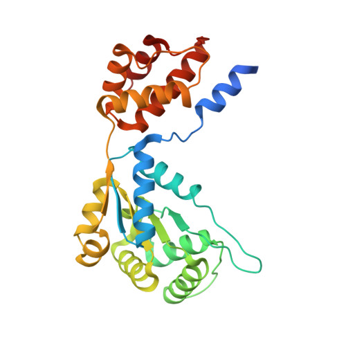
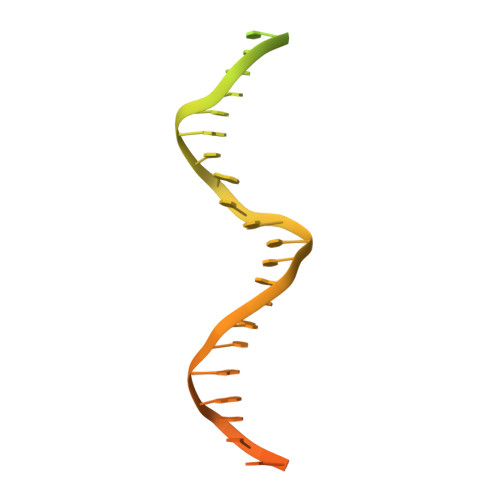
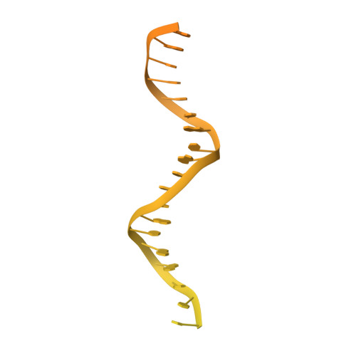
Conformational landscape of the type V-K CRISPR-associated transposon integration assembly.

Tenjo-Castano, F.Sofos, N.Stutzke, L.S.Temperini, P.Fuglsang, A.Pape, T.Mesa, P.Montoya, G.

(2024) Mol Cell 84: 2353

  • DOI: https://doi.org/10.1016/j.molcel.2024.05.005
  • Primary Citation Related Structures: 
    8AXA, 8AXB, 8RDU, 8RKT, 8RKU, 8RKV

  • PubMed Abstract: 

    CRISPR-associated transposons (CASTs) are mobile genetic elements that co-opt CRISPR-Cas systems for RNA-guided DNA transposition. CASTs integrate large DNA cargos into the attachment (att) site independently of homology-directed repair and thus hold promise for eukaryotic genome engineering. However, the functional diversity and complexity of CASTs hinder an understanding of their mechanisms. Here, we present the high-resolution cryoelectron microscopy (cryo-EM) structure of the reconstituted ∼1 MDa post-transposition complex of the type V-K CAST, together with different assembly intermediates and diverse TnsC filament lengths, thus enabling the recapitulation of the integration complex formation. The results of mutagenesis experiments probing the roles of specific residues and TnsB-binding sites show that transposition activity can be enhanced and suggest that the distance between the PAM and att sites is determined by the lengths of the TnsB C terminus and the TnsC filament. This singular model of RNA-guided transposition provides a foundation for repurposing the system for genome-editing applications.


  • Organizational Affiliation
    • Structural Molecular Biology Group, Novo Nordisk Foundation Centre for Protein Research, Faculty of Health and Medical Sciences, University of Copenhagen, Blegdamsvej 3B, 2200 Copenhagen, Denmark.

Macromolecule Content 

  • Total Structure Weight: 508.83 kDa 
  • Atom Count: 30,726 
  • Modeled Residue Count: 3,638 
  • Deposited Residue Count: 4,065 
  • Unique protein chains: 1
  • Unique nucleic acid chains: 2

Macromolecules


Find similar proteins by:|  3D Structure
Entity ID: 3
MoleculeChains  Sequence LengthOrganismDetailsImage
ShTnsC276Scytonema hofmanniiMutation(s): 0 
UniProt
Find proteins for A0A8J0PCL3 (Scytonema hofmannii)
Explore A0A8J0PCL3 
Go to UniProtKB:  A0A8J0PCL3
Entity Groups
Sequence Clusters30% Identity50% Identity70% Identity90% Identity95% Identity100% Identity
UniProt GroupA0A8J0PCL3
Sequence Annotations
Expand
Reference Sequence
Find similar nucleic acids by:  (by identity cutoff) 
Entity ID: 1
MoleculeChains LengthOrganismImage
Non-target strand - LEA [auth 2]68Scytonema hofmannii
Sequence Annotations
Expand
Reference Sequence
Find similar nucleic acids by:  (by identity cutoff) 
Entity ID: 2
MoleculeChains LengthOrganismImage
Target strand - LEB [auth 3]133Scytonema hofmannii
Sequence Annotations
Expand
Reference Sequence

Small Molecules

Ligands 2 Unique
IDChains Name / Formula / InChI Key2D Diagram3D Interactions
ATP
(Subject of Investigation/LOI)

Query on ATP


Download:Ideal Coordinates CCD File
AA [auth I]
CA [auth J]
EA [auth K]
GA [auth L]
IA [auth M]
AA [auth I],
CA [auth J],
EA [auth K],
GA [auth L],
IA [auth M],
KA [auth N],
MA [auth O],
OA [auth P],
R [auth D],
RA [auth Q],
S [auth E],
U [auth F],
W [auth G],
Y [auth H]
ADENOSINE-5'-TRIPHOSPHATE
C10 H16 N5 O13 P3
ZKHQWZAMYRWXGA-KQYNXXCUSA-N
MG
(Subject of Investigation/LOI)

Query on MG


Download:Ideal Coordinates CCD File
BA [auth I]
DA [auth J]
FA [auth K]
HA [auth L]
JA [auth M]
BA [auth I],
DA [auth J],
FA [auth K],
HA [auth L],
JA [auth M],
LA [auth N],
NA [auth O],
PA [auth P],
Q [auth D],
QA [auth Q],
T [auth E],
V [auth F],
X [auth G],
Z [auth H]
MAGNESIUM ION
Mg
JLVVSXFLKOJNIY-UHFFFAOYSA-N

Experimental Data & Validation

Experimental Data

  • Method: ELECTRON MICROSCOPY
  • Resolution: 2.30 Å
  • Aggregation State: PARTICLE 
  • Reconstruction Method: SINGLE PARTICLE 
EM Software:
TaskSoftware PackageVersion
RECONSTRUCTIONcryoSPARC4.4
MODEL REFINEMENTPHENIX
MODEL REFINEMENTISOLDE
MODEL REFINEMENTCoot

Structure Validation

View Full Validation Report


Entry History 

& Funding Information

Deposition Data


Funding OrganizationLocationGrant Number
Novo Nordisk FoundationDenmarkNNF14CC0001
European Research Council (ERC)European Union101096548

Revision History  (Full details and data files)

  • Version 1.0: 2024-06-19
    Type: Initial release
  • Version 1.1: 2024-07-03
    Changes: Data collection, Database references