8RKE | pdb_00008rke

Crystal structure of the complete N-terminal region of human ZP2 (hZP2-N1N2N3)


Experimental Data Snapshot

  • Method: X-RAY DIFFRACTION
  • Resolution: 2.70 Å
  • R-Value Free: 
    0.258 (Depositor), 0.260 (DCC) 
  • R-Value Work: 
    0.229 (Depositor), 0.230 (DCC) 
  • R-Value Observed: 
    0.230 (Depositor) 

wwPDB Validation 3D Report Full Report

Validation slider image for 8RKE

Ligand Structure Quality Assessment 


This is version 1.2 of the entry. See complete history

Literature

ZP2 cleavage blocks polyspermy by modulating the architecture of the egg coat.

Nishio, S.Emori, C.Wiseman, B.Fahrenkamp, D.Dioguardi, E.Zamora-Caballero, S.Bokhove, M.Han, L.Stsiapanava, A.Algarra, B.Lu, Y.Kodani, M.Bainbridge, R.E.Komondor, K.M.Carlson, A.E.Landreh, M.de Sanctis, D.Yasumasu, S.Ikawa, M.Jovine, L.

(2024) Cell 187: 1440-1459.e24

  • DOI: https://doi.org/10.1016/j.cell.2024.02.013
  • Primary Citation Related Structures: 
    8BQU, 8RKE, 8RKF, 8RKG, 8RKH, 8RKI

  • PubMed Abstract: 

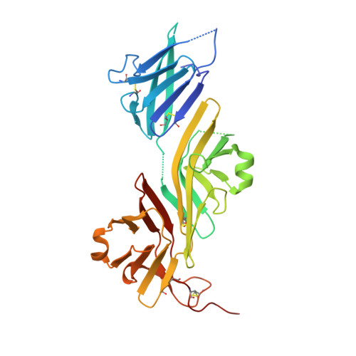
    Following the fertilization of an egg by a single sperm, the egg coat or zona pellucida (ZP) hardens and polyspermy is irreversibly blocked. These events are associated with the cleavage of the N-terminal region (NTR) of glycoprotein ZP2, a major subunit of ZP filaments. ZP2 processing is thought to inactivate sperm binding to the ZP, but its molecular consequences and connection with ZP hardening are unknown. Biochemical and structural studies show that cleavage of ZP2 triggers its oligomerization. Moreover, the structure of a native vertebrate egg coat filament, combined with AlphaFold predictions of human ZP polymers, reveals that two protofilaments consisting of type I (ZP3) and type II (ZP1/ZP2/ZP4) components interlock into a left-handed double helix from which the NTRs of type II subunits protrude. Together, these data suggest that oligomerization of cleaved ZP2 NTRs extensively cross-links ZP filaments, rigidifying the egg coat and making it physically impenetrable to sperm.


  • Organizational Affiliation
    • Department of Biosciences and Nutrition, Karolinska Institutet, Huddinge, Sweden.

Macromolecule Content 

  • Total Structure Weight: 115.61 kDa 
  • Atom Count: 6,886 
  • Modeled Residue Count: 845 
  • Deposited Residue Count: 1,005 
  • Unique protein chains: 1

Macromolecules

Find similar proteins by:|  3D Structure
Entity ID: 1
MoleculeChains  Sequence LengthOrganismDetailsImage
Zona pellucida sperm-binding protein 2
A, B, C
335Homo sapiensMutation(s): 0 
Gene Names: ZP2ZPA
UniProt & NIH Common Fund Data Resources
Find proteins for Q05996 (Homo sapiens)
Explore Q05996 
Go to UniProtKB:  Q05996
PHAROS:  Q05996
GTEx:  ENSG00000103310 
Entity Groups
Sequence Clusters30% Identity50% Identity70% Identity90% Identity95% Identity100% Identity
UniProt GroupQ05996
Glycosylation
Glycosylation Sites: 1Go to GlyGen: Q05996-1
Sequence Annotations
Expand
Reference Sequence

Oligosaccharides

Help  
Entity ID: 2
MoleculeChains Length2D Diagram GlycosylationD Interactions
alpha-D-mannopyranose-(1-3)-alpha-D-mannopyranose-(1-6)-[alpha-D-mannopyranose-(1-3)]beta-D-mannopyranose-(1-4)-2-acetamido-2-deoxy-beta-D-glucopyranose-(1-4)-2-acetamido-2-deoxy-beta-D-glucopyranose
D
6N-Glycosylation
Glycosylation Resources
GlyTouCan: G09724ZC
GlyCosmos: G09724ZC
GlyGen: G09724ZC
Entity ID: 3
MoleculeChains Length2D Diagram GlycosylationD Interactions
alpha-D-mannopyranose-(1-3)-[alpha-D-mannopyranose-(1-6)]alpha-D-mannopyranose-(1-6)-[alpha-D-mannopyranose-(1-3)]beta-D-mannopyranose-(1-4)-2-acetamido-2-deoxy-beta-D-glucopyranose-(1-4)-2-acetamido-2-deoxy-beta-D-glucopyranose
E
7N-Glycosylation
Glycosylation Resources
GlyTouCan: G55220VL
GlyCosmos: G55220VL
GlyGen: G55220VL

Experimental Data & Validation

Experimental Data

  • Method: X-RAY DIFFRACTION
  • Resolution: 2.70 Å
  • R-Value Free:  0.258 (Depositor), 0.260 (DCC) 
  • R-Value Work:  0.229 (Depositor), 0.230 (DCC) 
  • R-Value Observed: 0.230 (Depositor) 
Space Group: C 1 2 1
Unit Cell:
Length ( Å )Angle ( ˚ )
a = 147.68α = 90
b = 97.77β = 94.803
c = 104.33γ = 90
Software Package:
Software NamePurpose
XDSdata reduction
XSCALEdata scaling
SHARPphasing
PHENIXmodel building
Cootmodel building
PHENIXrefinement

Structure Validation

View Full Validation Report


Ligand Structure Quality Assessment 


Entry History 

& Funding Information

Deposition Data


Funding OrganizationLocationGrant Number
Swedish Research CouncilSweden2016-03999
Swedish Research CouncilSweden2020-04936
Knut and Alice Wallenberg FoundationSweden2018.0042

Revision History  (Full details and data files)

  • Version 1.0: 2024-03-13
    Type: Initial release
  • Version 1.1: 2024-03-27
    Changes: Database references
  • Version 1.2: 2024-11-13
    Changes: Structure summary