8R0P | pdb_00008r0p

Cryo-EM structure of the microbial rhodopsin CryoR2 at pH 8.0 in detergent


Experimental Data Snapshot

  • Method: ELECTRON MICROSCOPY
  • Resolution: 2.44 Å
  • Aggregation State: PARTICLE 
  • Reconstruction Method: SINGLE PARTICLE 

wwPDB Validation 3D Report Full Report

Validation slider image for 8R0P

This is version 1.1 of the entry. See complete history

Literature

CryoRhodopsins: A comprehensive characterization of a group of microbial rhodopsins from cold environments.

Lamm, G.H.U.Marin, E.Alekseev, A.Schellbach, A.V.Stetsenko, A.Haro-Moreno, J.M.Bourenkov, G.Borshchevskiy, V.Asido, M.Agthe, M.Engilberge, S.Rose, S.L.Caramello, N.Royant, A.Schneider, T.R.Bateman, A.Mager, T.Moser, T.Rodriguez-Valera, F.Wachtveitl, J.Guskov, A.Kovalev, K.

(2025) Sci Adv 11: eadv1015-eadv1015

  • DOI: https://doi.org/10.1126/sciadv.adv1015
  • Primary Citation Related Structures: 
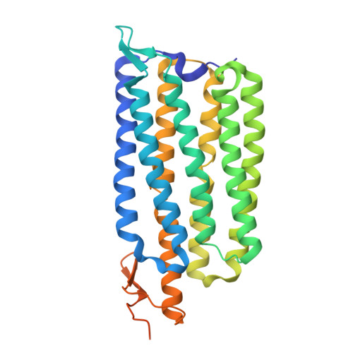
    8R0K, 8R0L, 8R0M, 8R0N, 8R0O, 8R0P

  • PubMed Abstract: 

    Microbial rhodopsins are omnipresent on Earth; however, the vast majority of them remain uncharacterized. Here, we describe a rhodopsin group found in microorganisms from cold environments, such as glaciers, denoted as CryoRhodopsins (CryoRs). A distinguishing feature of the group is the presence of a buried arginine residue close to the cytoplasmic face. Combining single-particle cryo-electron microscopy and x-ray crystallography with rhodopsin activation by light, we demonstrate that the arginine stabilizes an ultraviolet (UV)-absorbing intermediate of an extremely slow CryoRhodopsin photocycle. Together with extensive spectroscopic characterization, our investigations on CryoR1 and CryoR2 proteins reveal mechanisms of photoswitching in the identified group. Our data suggest that CryoRs are sensors for UV irradiation and are also capable of inward proton translocation modulated by UV light.


  • Organizational Affiliation
    • Institute of Physical and Theoretical Chemistry, Goethe University Frankfurt, 60438 Frankfurt am Main, Germany.

Macromolecule Content 

  • Total Structure Weight: 186.18 kDa 
  • Atom Count: 11,154 
  • Modeled Residue Count: 1,410 
  • Deposited Residue Count: 1,635 
  • Unique protein chains: 1

Macromolecules

Find similar proteins by:|  3D Structure
Entity ID: 1
MoleculeChains  Sequence LengthOrganismDetailsImage
microbial rhodopsin CryoR2
A, B, C, D, E
327Subtercola endophyticusMutation(s): 0 

Small Molecules

Ligands 2 Unique
IDChains Name / Formula / InChI Key2D Diagram3D Interactions
RET
(Subject of Investigation/LOI)

Query on RET


Download:Ideal Coordinates CCD File
CA [auth C],
KA [auth D],
M [auth A],
SA [auth E],
U [auth B]
RETINAL
C20 H28 O
NCYCYZXNIZJOKI-OVSJKPMPSA-N
LFA

Query on LFA


Download:Ideal Coordinates CCD File
AA [auth C]
BA [auth C]
DA [auth D]
EA [auth D]
F [auth A]
AA [auth C],
BA [auth C],
DA [auth D],
EA [auth D],
F [auth A],
FA [auth D],
G [auth A],
GA [auth D],
H [auth A],
HA [auth D],
I [auth A],
IA [auth D],
J [auth A],
JA [auth D],
K [auth A],
L [auth A],
LA [auth E],
MA [auth E],
N [auth B],
NA [auth E],
O [auth B],
OA [auth E],
P [auth B],
PA [auth E],
Q [auth B],
QA [auth E],
R [auth B],
RA [auth E],
S [auth B],
T [auth B],
V [auth C],
W [auth C],
X [auth C],
Y [auth C],
Z [auth C]
EICOSANE
C20 H42
CBFCDTFDPHXCNY-UHFFFAOYSA-N

Experimental Data & Validation

Experimental Data

  • Method: ELECTRON MICROSCOPY
  • Resolution: 2.44 Å
  • Aggregation State: PARTICLE 
  • Reconstruction Method: SINGLE PARTICLE 
EM Software:
TaskSoftware PackageVersion
RECONSTRUCTIONcryoSPARC4.1.2

Structure Validation

View Full Validation Report


Entry History 

& Funding Information

Deposition Data


Funding OrganizationLocationGrant Number
EIPOD fellowship under Marie Sklodowska-Curie Actions COFUNDGermany847543

Revision History  (Full details and data files)

  • Version 1.0: 2025-05-14
    Type: Initial release
  • Version 1.1: 2025-07-16
    Changes: Data collection, Database references